1,122
Views
12
CrossRef citations to date
0
Altmetric
Original Articles

Morphological and phylogenetic comparisons of Neodenticula seminae (Bacillariophyta) populations between the subarctic Pacific and the Gulf of St. Lawrence

, , , &
Pages 127-142 | Received 06 Apr 2009, Accepted 10 Sep 2009, Published online: 11 Jun 2010

Figures & data

Table 1. Strain designation, origin and accession numbers for Fragilariopsis strains from GenBank and Neodenticula seminae used in the phylogenetic analyses. Strains not mentioned here are found in Lundholm et al. (Citation2002).

Figs 1–3. Neodenticula seminae from the Gulf of St. Lawrence, Northwest Atlantic, light microscopy. . Straight ribbon-shaped colonies. . Coiled ribbon-shaped colony. . Solitary and paired cells with chloroplasts.
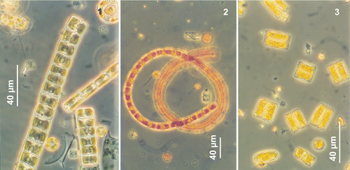
Figs 1–3. Neodenticula seminae from the Gulf of St. Lawrence, Northwest Atlantic, light microscopy. Fig. 1. Straight ribbon-shaped colonies. Fig. 2. Coiled ribbon-shaped colony. Fig. 3. Solitary and paired cells with chloroplasts.

Table 2. Morphometric data on Neodenticula seminae recorded from the subarctic Pacific, the Gulf of St. Lawrence and the literature.

Figs 4–11. Neodenticula seminae, light microscopy. . Valves of specimens from the Gulf of St. Lawrence () and subarctic Pacific () showing simple and vestigial-walled costae. . Valves of specimens from the subarctic Pacific showing the deck and basal ridges with foramina. . Phase contrast. . Brightfield. . Interference contrast.
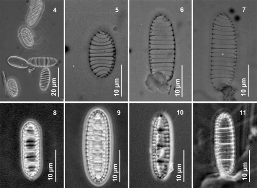
Figs 4–11. Neodenticula seminae, light microscopy. Figs 4–7, 11. Valves of specimens from the Gulf of St. Lawrence (Figs 4–7) and subarctic Pacific (Fig. 11) showing simple and vestigial-walled costae. Figs 8–10. Valves of specimens from the subarctic Pacific showing the deck and basal ridges with foramina. Figs 4, 8, 9, 11. Phase contrast. Figs 5–7. Brightfield. Fig. 10. Interference contrast.

Figs 12–21. Neodenticula seminae, electron microscopy (scanning electron microscopy unless indicated otherwise) of cultured () and natural () Gulf of St. Lawrence specimens and natural subarctic Pacific specimens (). . Whole frustule in girdle view. . Oblique view of valve showing full ornamentation on mantle (transmission electron microscopy). . Valve showing costae, areolate pattern and fibulate raphe system (transmission electron microscopy). . End pole of frustule showing raphe valve opposing non-raphe valve. . Broken frustule with hyaline girdle bands showing ligulate and antiligulate copulae. Note uninterrupted raphe fissure. . Series of girdle bands with ligulate copulae. . Valvocopula (arrow) adhering firmly to the valve. . Closed valvocopula. . Ligulate and antiligulate copulae at end of valve. . Open copula.
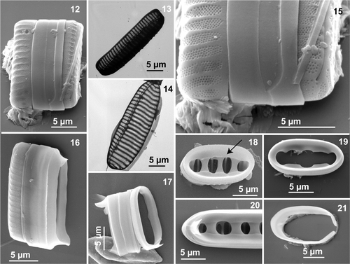
Figs 12–21. Neodenticula seminae, electron microscopy (scanning electron microscopy unless indicated otherwise) of cultured (Figs 12, 15, 16, 19, 21) and natural (Figs 13, 14) Gulf of St. Lawrence specimens and natural subarctic Pacific specimens (Figs 17, 18, 20). Fig. 12. Whole frustule in girdle view. Fig. 13. Oblique view of valve showing full ornamentation on mantle (transmission electron microscopy). Fig. 14. Valve showing costae, areolate pattern and fibulate raphe system (transmission electron microscopy). Fig. 15. End pole of frustule showing raphe valve opposing non-raphe valve. Fig. 16. Broken frustule with hyaline girdle bands showing ligulate and antiligulate copulae. Note uninterrupted raphe fissure. Fig. 17. Series of girdle bands with ligulate copulae. Fig. 18. Valvocopula (arrow) adhering firmly to the valve. Fig. 19. Closed valvocopula. Fig. 20. Ligulate and antiligulate copulae at end of valve. Fig. 21. Open copula.

Figs 22–33. Morphological variability in Neodenticula seminae under scanning electron microscopy. . External views of valves (. Natural specimens from the Gulf of St. Lawrence and, from subarctic Pacific). . Internal valve views showing vestigial-walled costae (arrow in ) (. Gulf of St. Lawrence, . Subarctic Pacific; . Cultured subarctic Pacific).
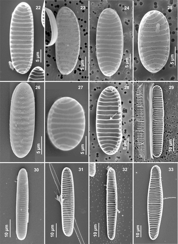
Figs 22–33. Morphological variability in Neodenticula seminae under scanning electron microscopy. Figs 22–27, 30. External views of valves (Figs 22–25, 27. Natural specimens from the Gulf of St. Lawrence and, Figs 26, 30 from subarctic Pacific). Figs 28, 29, 31–33. Internal valve views showing vestigial-walled costae (arrow in Fig. 28) (Figs 28, 29. Gulf of St. Lawrence, Fig. 31. Subarctic Pacific; Figs 32, 33. Cultured subarctic Pacific).

Figs 34–40. Neodenticula seminae, internal views, scanning electron microscopy. . Valve showing simple and vestigial-walled costae (arrow) (subarctic Pacific). . Whole valve (Gulf of St. Lawrence culture). . Apical end showing the raphe canal and curved apical costae (subarctic Pacific and Gulf of St. Lawrence cultures, respectively). . Valve with deck (arrow) and three basal ridges (asterisk) (subarctic Pacific). . Part of valve showing foramina (asterisk), simple costae (long arrow) and solid-walled costae (short arrow) connecting to basal ridges (subarctic Pacific). . Two valves with basal ridges and foramina (subarctic Pacific).
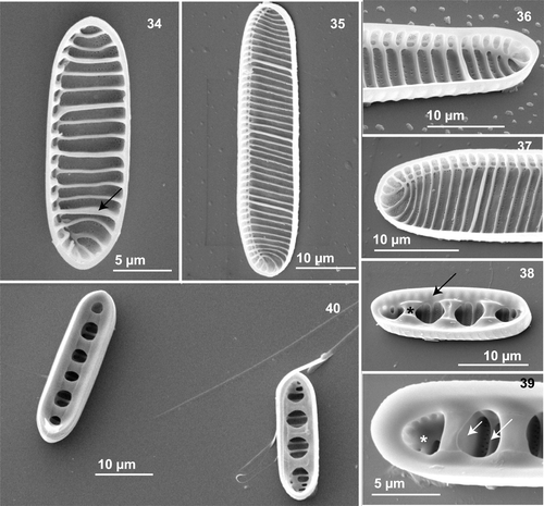
Figs 34–40. Neodenticula seminae, internal views, scanning electron microscopy. Fig. 34. Valve showing simple and vestigial-walled costae (arrow) (subarctic Pacific). Fig. 35. Whole valve (Gulf of St. Lawrence culture). Figs 36, 37. Apical end showing the raphe canal and curved apical costae (subarctic Pacific and Gulf of St. Lawrence cultures, respectively). Fig. 38. Valve with deck (arrow) and three basal ridges (asterisk) (subarctic Pacific). Fig. 39. Part of valve showing foramina (asterisk), simple costae (long arrow) and solid-walled costae (short arrow) connecting to basal ridges (subarctic Pacific). Fig. 40. Two valves with basal ridges and foramina (subarctic Pacific).

Figs 41–46. Neodenticula seminae, scanning electron microscopy. . Valve showing the external and continuous raphe fissure and full areolate pattern on mantle (Gulf of St. Lawrence). . External views of apex showing raphe ending and ornamentation on mantle (Gulf of St. Lawrence culture and natural material, respectively). Note the very gentle peeling off at the valve–mantle junction in . . External valve centre showing the uninterrupted raphe fissure. Note the fully developed ornamentation on mantle and the very scarce areolae on the valve surface (subarctic Pacific culture). . Internal view of apex (Gulf of St. Lawrence culture). . Apex showing the internal costae and the raphe canal (Gulf of St. Lawrence culture).
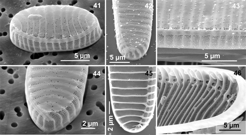
Figs 41–46. Neodenticula seminae, scanning electron microscopy. Fig. 41. Valve showing the external and continuous raphe fissure and full areolate pattern on mantle (Gulf of St. Lawrence). Figs 42, 44. External views of apex showing raphe ending and ornamentation on mantle (Gulf of St. Lawrence culture and natural material, respectively). Note the very gentle peeling off at the valve–mantle junction in Fig. 44. Fig. 43. External valve centre showing the uninterrupted raphe fissure. Note the fully developed ornamentation on mantle and the very scarce areolae on the valve surface (subarctic Pacific culture). Fig. 45. Internal view of apex (Gulf of St. Lawrence culture). Fig. 46. Apex showing the internal costae and the raphe canal (Gulf of St. Lawrence culture).

Figs 47–54. Electron microscopy of Neodenticula seminae. . External views, scanning electron microscopy (SEM). . Internal view, SEM. . transmission electron microscopy (TEM). . SEM, external views of apex showing the terminal raphe ending, curved virgae and scarce areolae on the valve surface. . Gulf of St. Lawrence culture, . subarctic Pacific culture. . TEM of almost circular valve with restricted but more complete areolation on the raphe-opposing margin (Gulf of St. Lawrence). . SEM showing spinulose structures (arrow) on the raphe-opposing margin (Gulf of St. Lawrence). . TEM of valve showing costae, reduced ornamentation and raphe canal (Gulf of St. Lawrence). . Part of the valve showing the external raphe fissure, virgae and ornamentation of the valve face and mantle (subarctic Pacific culture). . SEM, internal view of apex showing scarce areolae between costae (Gulf of St. Lawrence culture).
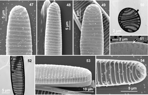
Figs 47–54. Electron microscopy of Neodenticula seminae. Figs 47–49, 51, 53. External views, scanning electron microscopy (SEM). Fig. 54. Internal view, SEM. Figs 50, 52. transmission electron microscopy (TEM). Figs 47–49. SEM, external views of apex showing the terminal raphe ending, curved virgae and scarce areolae on the valve surface. Figs 47, 49. Gulf of St. Lawrence culture, Fig. 48. subarctic Pacific culture. Fig. 50. TEM of almost circular valve with restricted but more complete areolation on the raphe-opposing margin (Gulf of St. Lawrence). Fig. 51. SEM showing spinulose structures (arrow) on the raphe-opposing margin (Gulf of St. Lawrence). Fig. 52. TEM of valve showing costae, reduced ornamentation and raphe canal (Gulf of St. Lawrence). Fig. 53. Part of the valve showing the external raphe fissure, virgae and ornamentation of the valve face and mantle (subarctic Pacific culture). Fig. 54. SEM, internal view of apex showing scarce areolae between costae (Gulf of St. Lawrence culture).

Fig. 55. Phylogeny of the Bacillariaceae inferred from maximum likelihood analysis of large subunit of the nuclear ribosomal DNA. Numbers show bootstrap values obtained by neighbor joining and maximum parsimony analyses, respectively.
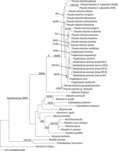
Fig. 55. Phylogeny of the Bacillariaceae inferred from maximum likelihood analysis of large subunit of the nuclear ribosomal DNA. Numbers show bootstrap values obtained by neighbor joining and maximum parsimony analyses, respectively.

Fig. 56. Phylogeny of Neodenticula seminae and Fragilariopsis species inferred from maximum likelihood analysis of internal transcribed spacer regions of the nuclear ribosomal DNA. Numbers show bootstrap values obtained by maximum parsimony, neighbor joining and maximum likelihood analyses, respectively.
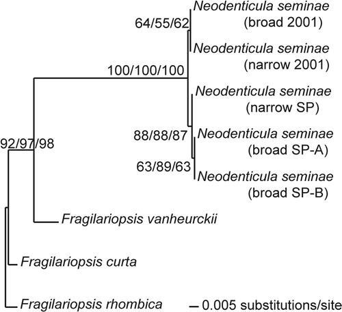
Fig. 56. Phylogeny of Neodenticula seminae and Fragilariopsis species inferred from maximum likelihood analysis of internal transcribed spacer regions of the nuclear ribosomal DNA. Numbers show bootstrap values obtained by maximum parsimony, neighbor joining and maximum likelihood analyses, respectively.

Reprints and Corporate Permissions

Please note: Selecting permissions does not provide access to the full text of the article, please see our help page How do I view content?

To request a reprint or corporate permissions for this article, please click on the relevant link below:

Academic Permissions

Please note: Selecting permissions does not provide access to the full text of the article, please see our help page How do I view content?

Obtain permissions instantly via Rightslink by clicking on the button below:

If you are unable to obtain permissions via Rightslink, please complete and submit this Permissions form. For more information, please visit our Permissions help page.