Figures & data
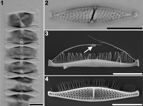
Figs 1–4. Plagiogrammopsis vanheurckii mature cells. Strain s0435, light microscopy () and strain s0432, scanning electron microscopy (). . Chain of living cells. . Cleaned process valve. . External view of pilus valve. Arrow indicates wing on pilus. . Internal view of process valve. Scale bars: 10 µm.
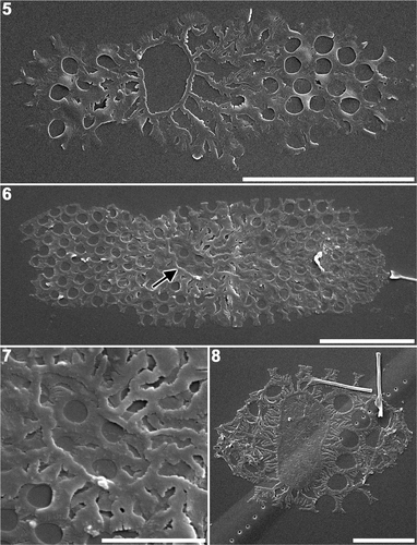
Figs 5–8. Plagiogrammopsis vanheurckii, strain s0433 () and strain s0435 (). Early stages of valve morphogenesis, scanning electron microscopy. . Valve with a distinct annulus, in which silica deposition has not commenced. . Pair of sibling valves showing different stages of development. Arrow indicates annulus of younger valve. . Enlarged view of the annulus shown in . . Valve with distinct annulus, in which silica deposition is nearly complete. Scale bars: 5 µm () and 2 µm ().
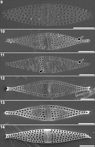
Figs 9–14. Plagiogrammopsis vanheurckii, strain s0432. Middle stages of valve morphogenesis, external view, scanning electron microscopy. . Silica deposition nearly completed inside annulus. . Plain disc at centre. Note presence of pseudoseptum beneath the disk. Arrow indicates row of base of marginal spines. . Fascia at centre. Arrow indicates base of marginal spines. Arrowhead indicates small areola where rimoportula will be formed. . Apex formation. Note distinct row of marginal spines. . Valve margin thickening. . Fully developed valve face and mantle, but apices not yet formed. Scale bars: 5 µm.
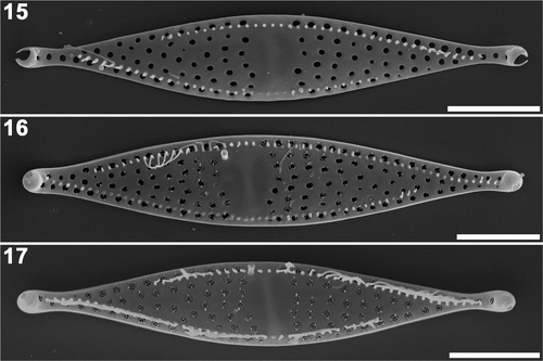
Figs 15–17. Plagiogrammopsis vanheurckii, strain s0434 () and strain s0432 (). Final stages of valve morphogenesis, external view, scanning electron microscopy. . Valve apex formation, ocelluli still unformed (strain s0434). . Almost mature valve during cribrum formation (strain s0432). . Mature valve (strain s0434). Scale bars: 5 µm.
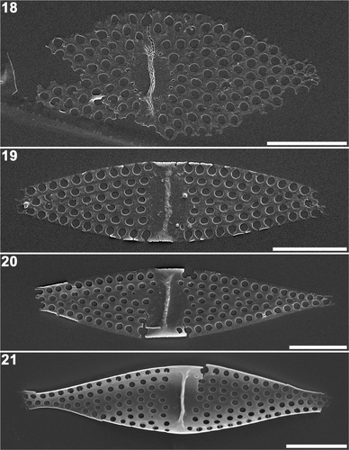
Figs 18–21. Plagiogrammopsis vanheurckii, strain s0433 () and strain s0432 (). Middle stages of valve morphogenesis, internal view, scanning electron microscopy. . Valve during radial expansion with pseudoseptum on the central disc. . Marginal thickening started around fascia. Note the valve margin is incomplete. . Lanceolate valve outline complete. . Three-dimensional valve framework complete. Note rimoportula on right hand side of pseudoseptum. Scale bars: 5 µm.
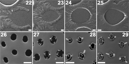
Figs 22–29. Plagiogrammopsis vanheurckii, strain s0432. Formation of areolae () and cribra (), scanning electron microscopy. Valve centre lies on the left side in . . Centrifugal extension of two semi-circular arms. . Tips of arms fuse at opposite side to form areola. . Thickening starts at fused site. . Thickening proceeds centripetally. . Bases of cribra seen as knobs. . Centripetal cribrum formation. . Mature cribra. Scale bars: 0.1 () and 0.5 µm ().
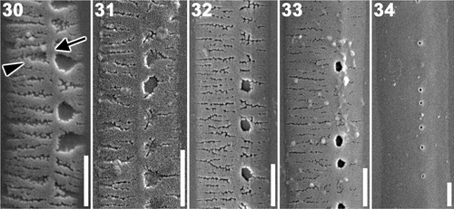
Figs 30–34. Plagiogrammopsis vanheurckii, strain s0432. Band formation, scanning electron microscopy. . Early stages of band formation comprise longitudinal (arrow) and transverse (arrowhead) ribs. . Middle stage. . Final stage. . Mature band. Scale bars: 0.5 µm.
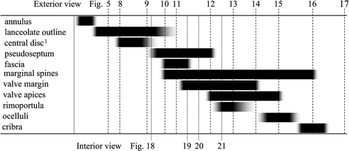
Fig. 35. Diagram of formation order of valve elements during valve morphogenesis in Plagiogrammopsis vanheurckii. 1Annulus becomes disc when silica deposition inside annulus is completed.