786
Views
9
CrossRef citations to date
0
Altmetric
Original Articles

Climaconeis species (Bacillariophyceae: Berkeleyaceae) from western Pacific islands, including C. petersonii sp. nov. and C. guamensis sp. nov., with emphasis on the plastids

, &
Pages 293-307 | Received 04 Nov 2009, Accepted 01 May 2010, Published online: 26 Aug 2010

Figures & data

Figs 1–4. Drawings of Climaconeis plastids from the literature. . Mereschkowsky's (Citation1901) Okedenia. . O. scopulorum. . O. scopulorum var. fasciculata. . O. inflexa. . Cox's (Citation1979) Navicula scopulorum B. [. reprinted with permission of the British Phycological Society.]

Figs 1–4. Drawings of Climaconeis plastids from the literature. Figs 1–3. Mereschkowsky's (Citation1901) Okedenia. Fig. 1. O. scopulorum. Fig. 2. O. scopulorum var. fasciculata. Fig. 3. O. inflexa. Fig. 4. Cox's (Citation1979) Navicula scopulorum B. [Fig. 4. reprinted with permission of the British Phycological Society.]

Table 1.  Collection sites for listed records.

Figs 5–18. Climaconeis plastids, freshly collected living cells, from Guam except as noted. . C. undulata. . Valve views, the cell in 5 showing some plastids possibly joined by pyrenoids (arrows). . Oblique view of small cell. . Girdle view showing pyrenoids. . C. petersonii. . Half of cell in valve view, composite image. . Central portion in girdle view, showing the conspicuous pyrenoids. . C. lorenzii from Palau, photographed after 24 h transport; arrowhead points to craticular bar. . C. silvae valve views. . Half cells showing concentration of plastids in the central area, plastids at an oblique angle in 12. . Details of plastids at several angles in valve views of cells. . C. riddleae (GU44Y-13) and enlargement to show striae. . Cells from GU44Y-13, probably C. riddleae but possibly C. guamensis, displaying pyrenoids in valve and girdle view (py). Scale bar for all = 20 µm.

Figs 5–18. Climaconeis plastids, freshly collected living cells, from Guam except as noted. Figs 5–8. C. undulata. Fig. 5–6. Valve views, the cell in 5 showing some plastids possibly joined by pyrenoids (arrows). Fig. 7. Oblique view of small cell. Fig. 8. Girdle view showing pyrenoids. Figs 9–10. C. petersonii. Fig. 9. Half of cell in valve view, composite image. Fig. 10. Central portion in girdle view, showing the conspicuous pyrenoids. Fig. 11. C. lorenzii from Palau, photographed after 24 h transport; arrowhead points to craticular bar. Figs 12–15. C. silvae valve views. Figs 12–13. Half cells showing concentration of plastids in the central area, plastids at an oblique angle in 12. Figs 14–15. Details of plastids at several angles in valve views of cells. Figs 16–17. C. riddleae (GU44Y-13) and enlargement to show striae. Fig. 18. Cells from GU44Y-13, probably C. riddleae but possibly C. guamensis, displaying pyrenoids in valve and girdle view (py). Scale bar for all = 20 µm.

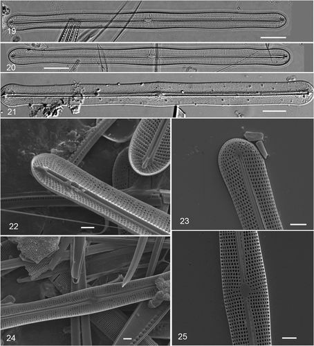
Figs 19–25. Climaconeis undulata acid-cleaned valves. . Two valves showing radiating stria pattern; field-collected material (GU16V), DIC. . Valve of Navicula scopulorum var. triundulata; slide N11/17 in Hustedt collection, prepared by Hustedt from Meister's material, photo H52078-3a courtesy of AWI (Citation2009) (DIC). . SEM of cultured cells. . Internal apex showing helictoglossa. . External apex. . Internal central area. . External central area. Scale bars: , 10 µm; 22–25 = 2 µm.

Figs 19–25. Climaconeis undulata acid-cleaned valves. Figs 19–20. Two valves showing radiating stria pattern; field-collected material (GU16V), DIC. Fig. 21. Valve of Navicula scopulorum var. triundulata; slide N11/17 in Hustedt collection, prepared by Hustedt from Meister's material, photo H52078-3a courtesy of AWI (Citation2009) (DIC). Figs 22–25. SEM of cultured cells. Fig. 22. Internal apex showing helictoglossa. Fig. 23. External apex. Fig. 24. Internal central area. Fig. 25. External central area. Scale bars: Fig. 19–21, 10 µm; 22–25 = 2 µm.

Figs 26–35. . Climaconeis petersonii acid-cleaned valves, field collected material. . Type specimen, GU55B-4, slide 2. DIC. . Details of central area and apex of type specimen. DIC. . Internal and external central areas, SEM. . External and internal apices, the latter showing the helictoglossa. SEM. . Climaconeis lorenzii acid-cleaned valve from Yap (Y26C). . Frustule showing deflected central raphe endings. . Valvocopula with craticular bars. . Climaconeis silvae, portion of acid-cleaned valve; field-collected material, GU44I-2, DIC. Scale bars: = 10 µm; = 2 µm.

Figs 26–35. Figs 26–32. Climaconeis petersonii acid-cleaned valves, field collected material. Fig. 26. Type specimen, GU55B-4, slide 2. DIC. Figs 27–28. Details of central area and apex of type specimen. DIC. Figs 29–30. Internal and external central areas, SEM. Figs 31–32. External and internal apices, the latter showing the helictoglossa. SEM. Figs 33–34. Climaconeis lorenzii acid-cleaned valve from Yap (Y26C). Fig. 33. Frustule showing deflected central raphe endings. Fig. 34. Valvocopula with craticular bars. Fig. 35. Climaconeis silvae, portion of acid-cleaned valve; field-collected material, GU44I-2, DIC. Scale bars: Figs 26–28, 33–35 = 10 µm; Figs 29–32 = 2 µm.

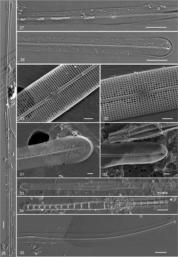
Table 2.  Characteristics of selected Climaconeis species. Data from literature and present study.

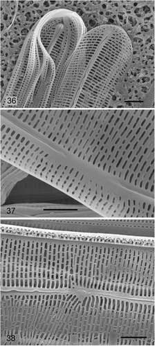
Figs 36–38. Climaconeis silvae SEM of cells cultured from Guam collection. . Internal and external apices and girdle bands. . Internal central area. . External central area. Scale bars = 2 µm.

Figs 36–38. Climaconeis silvae SEM of cells cultured from Guam collection. Fig. 36. Internal and external apices and girdle bands. Figs 37. Internal central area. Fig. 38. External central area. Scale bars = 2 µm.

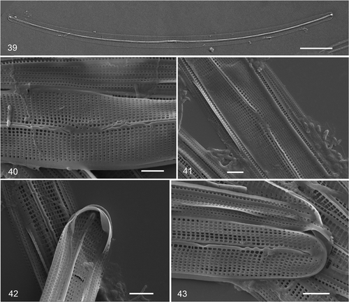
Figs 39–43. Climaconeis riddleae from Guam. . Acid-cleaned valve from GU44Y-13. . SEMs from isolate ECT 3722. . External and internal views of central area. . Internal and external views of apex. Scale bars: = 10 µm; = 2 µm.

Figs 39–43. Climaconeis riddleae from Guam. Fig. 39. Acid-cleaned valve from GU44Y-13. Figs 40–43. SEMs from isolate ECT 3722. Figs 40–41. External and internal views of central area. Figs 42–43. Internal and external views of apex. Scale bars: Fig. 39 = 10 µm; Figs 40–43 = 2 µm.

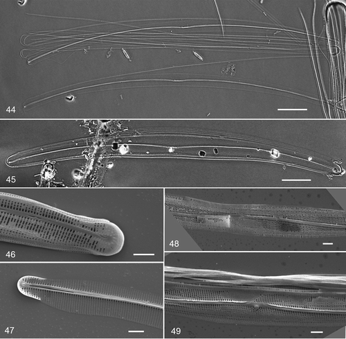
Figs 44–49. Climaconeis guamensis acid-cleaned valves. . Holotype specimen, GU44Y-13, slide 1. DIC. . Specimen from isotype slide, isolate ECT 3737. DIC. . SEM of cultured cells, isolate ECT 3737. . Internal and external views of apex. . External and internal views of central area. Scale bars: = 10 µm; = 2 µm.

Figs 44–49. Climaconeis guamensis acid-cleaned valves. Fig. 44. Holotype specimen, GU44Y-13, slide 1. DIC. Fig. 45. Specimen from isotype slide, isolate ECT 3737. DIC. Figs 46–49. SEM of cultured cells, isolate ECT 3737. Figs 46–47. Internal and external views of apex. Figs 48–49. External and internal views of central area. Scale bars: Figs 44–45 = 10 µm; Figs 46–49 = 2 µm.

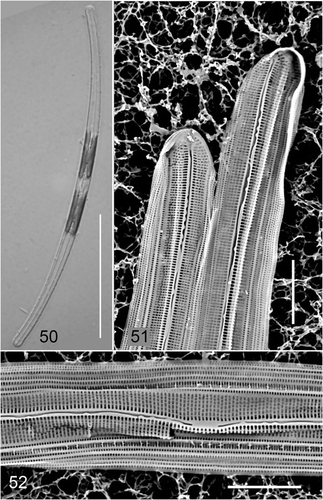
Figs 50–52. Climaconeis guamensis from GU54A-F. . Live cell. . SEM from whole mount of same material. Scale bars: = 50 µm; = 5 µm.

Figs 50–52. Climaconeis guamensis from GU54A-F. Fig. 50. Live cell. Figs 51–52. SEM from whole mount of same material. Scale bars: Fig. 50 = 50 µm; Figs 51–52 = 5 µm.

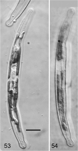
Figs 53–54. Climaconeis inflexa from Guam. . Cell with plastids in focus. . Cell with striae in focus. Scale bar = 10 µm.

Figs 53–54. Climaconeis inflexa from Guam. Fig. 53. Cell with plastids in focus. Fig. 54. Cell with striae in focus. Scale bar = 10 µm.

Reprints and Corporate Permissions

Please note: Selecting permissions does not provide access to the full text of the article, please see our help page How do I view content?

To request a reprint or corporate permissions for this article, please click on the relevant link below:

Academic Permissions

Please note: Selecting permissions does not provide access to the full text of the article, please see our help page How do I view content?

Obtain permissions instantly via Rightslink by clicking on the button below:

If you are unable to obtain permissions via Rightslink, please complete and submit this Permissions form. For more information, please visit our Permissions help page.