Figures & data
Fig. 1. Features of Rivularia colonies. (a) Diagram of a vertical section through a mature Rivularia colony showing orientation of the trichomes and two bands of calcification. (b) Sampling block for biovolume and CaCO3 content. (c) Part of the Rivularia colony showing trichomes enclosed by a branched mucilaginous sheath. (d) Vertical view of a Rivularia colony showing trichomes (filled circles) surrounded by firm mucilaginous sheaths (stippled) enclosed in highly hydrated and diffluent mucilage in which calcification occurs (white).
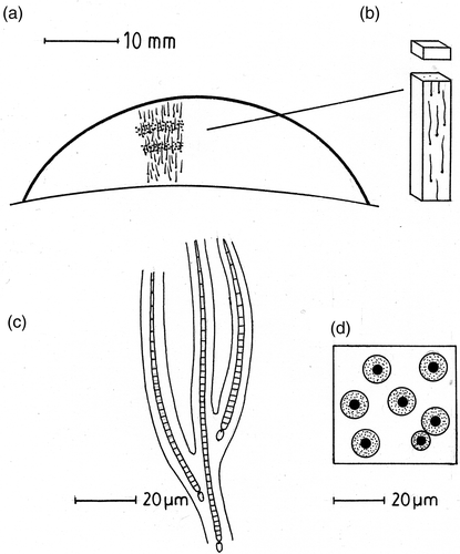
Table 1. Water chemical composition at the two sampling sites. Measurements, excluding pH and temperature, in mmol l−1 unless otherwise stated.
Fig. 2. Trichome density in Rivularia colonies. (a) Number of trichomes per mm2 of surface vs depth within colony (mm). (b) Trichome relative biovolume vs depth. Filled circles, R. haematites colonies, open circles, R. biasolettiana.
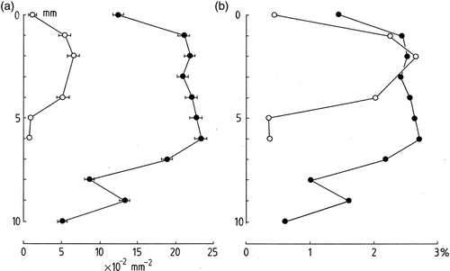
Fig. 3. Calcium carbonate content (vol. %) of Rivularia with depth. (a) R. haematites. (b) R. biasolettiana. Accurate values are not available for deepest parts of colony owing to breakdown of mucilage. Measurements averaged from duplicate samples taken from single colony.
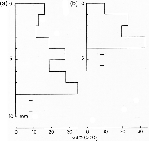
Fig. 4. Light absorption in Rivularia colonies as a function of depth. Filled circles, R. haematites; open circles, R. biasolettiana. Linear regression lines for R. haematites (full) and R. biasolettiana (broken) are included.
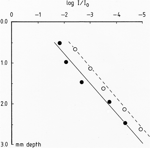
Fig. 5. Dissolved oxygen concentration in a R. haematites colony. Porphyrin planar optode at a surface irradiance of 500 µmol m−2 s−1. The thin black line indicates the colony surface. Silhouette of colony section shown below.
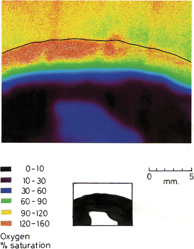
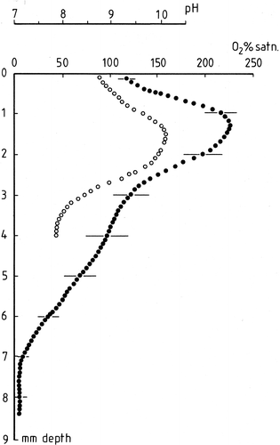
Fig. 6. Oxygen profile in a R. biasolettiana colony. Mean of three replicate runs showing standard errors at 1 mm intervals. Ordinate gives distance from colony surface in mm. For oxygen, only alternate datapoints are shown on this figure; the original resolution was 50 µm. Filled circles: microelectrode % oxygen. Open circles: pH at a resolution of 100 µm. Irradiance 500 µmol m−2 s−1 for both measurements.