Figures & data
Table 1. Amplification and sequencing primers used in this study to obtain rbcL and nuSSU sequences for Verrucariaceae photobionts. The abbreviations ‘amp’ and ‘seq’ indicate primers used for amplification and sequencing, respectively.
Fig. 1. Phylogenetic placement of the xanthophycean photobionts of Verrucariaceae. The tree was obtained using a maximum-likelihood analysis of a two-gene dataset (rbcL–nuSSU). Lichenized algal strains are indicated in parentheses with the name of the corresponding lichen-forming fungus and its collection number. Bootstrap values >70% are indicated above or below the branches.
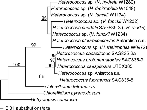
Fig. 2. Phylogenetic placement of chlorophyte photobionts of Verrucariaceae. The tree was obtained using a maximum-likelihood analysis of a two-gene dataset (rbcL–nuSSU). Lichenized algal strains are indicated by the addition, in parentheses, of the name of the corresponding lichen-forming fungus. They all belong to the family Verrucariaceae, except for Cladonia grayi, Icmadophila ericetorum, Lobaria linita, Racodium rupestre, Stictis urceolatum and Xanthoria parietina. For the Verrucariaceae, the collection numbers follow the fungal names. Bootstrap values >70% are indicated above or below the branches. Photobionts with identical sequences are represented by a single taxon in the phylogeny with the names and collection numbers of each lichen indicated.
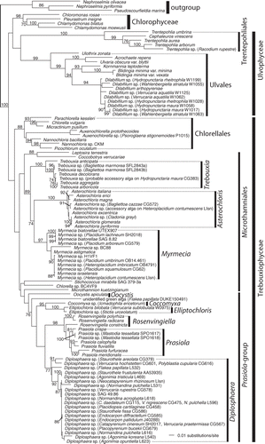
Fig. 3. Phylogenetic placement of the photobionts of Verrucariaceae related to the Prasiola-group. The tree was obtained using a maximum-likelihood analysis of a two-gene dataset (rbcL–nuSSU). Lichenized algal strains are indicated by the addition, in parentheses, of the name of the corresponding lichen-forming fungus. They all belong to the family Verrucariaceae, except for Icmadophila ericetorum, Lobaria pulmonaria, Phlyctis argena and Stictis urceolatum. For the Verrucariaceae, the collection numbers follow the fungal names. Bootstrap values >70% are indicated above or below the branches, except for values of 100%, which are indicated by a star. Photobionts with identical sequences are represented by a single taxon in the phylogeny with the names and collection numbers of each lichen indicated.
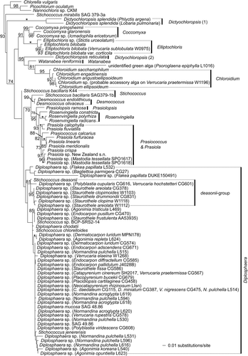
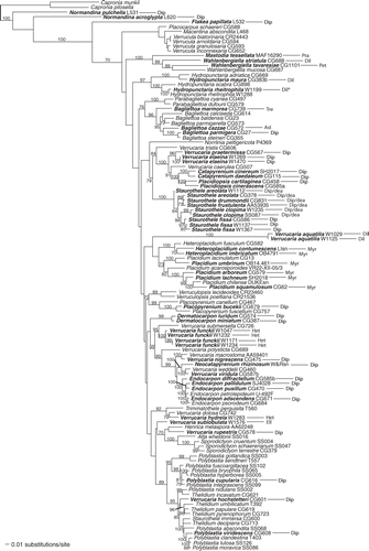
Fig. 4. Distribution of photobionts across a phylogeny of the Verrucariaceae obtained using a maximum-likelihood analysis of a four-gene dataset (RPB1–nuLSU–nuSSU–mtSSU). Lichen specimens for which molecular data are available for the photobionts are indicated in bold. For these specimens, the identity of the photobiont is shown after the taxon label using the following abbreviations: Ast = Asterochloris, Dil = Dilabifilum, Dip = Diplosphaera, Dip/dea = Diplosphaera deasonii-group (Stichococcus aff. mirabilis), Ell = Elliptochloris, Het = Heterococcus, Myr = Myrmecia, Pet = Petroderma maculiforme, Pra = Prasiola, Tre = Trebouxia. *For Hydropunctaria rheitrophila, two other specimens not included in this tree (W1048 and W0972) were associated with Heterococcus sp. (see ).
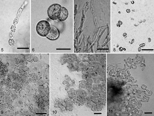
Figs 5–11. Light photomicrographs of algae isolated from crustose lichens of the family Verrucariaceae. 5. Filamentous growth in young culture of Heterococcus sp. isolated from Verrucaria funckii and cultured on 1.5% BBM-agar (BM H. Thüs W1174). 6. Cell aggregates of Heterococcus sp. isolated from V. funckii and cultured in liquid BBM medium (BM H. Thüs W1174). 7. Dilabifilum sp. isolated from V. aquatilis (BM H. Thüs W1125). 8. Chloroidium sp. isolated from V. praetermissa (BM H. Thüs W1198). 9. Elliptochloris bilobata isolated from V. sublobulata (BM H. Thüs W0975). 10. Diplosphaera sp. isolated from V. elaeina (BM H. Thüs W1269). 11. Stichococcus cf. mirabilis isolated from Staurothele clopima (BM H. Thüs W1118). Scale bars: 10 µm.