Figures & data
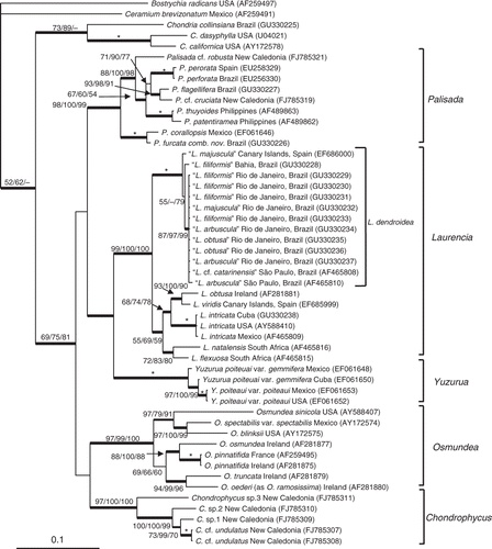
Fig. 1. Consensus tree derived from Bayesian analyses of rbcL sequences. The posterior probabilities (when > 95%) are shown as thicker branches. Bootstrap supports for MP/NJ (2000 replicates)/ML (100 replicates) are shown at the nodes; – indicates lack of bootstrap support; * indicates bootstrap support = 100%.
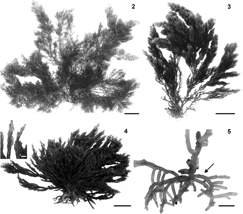
Figs 2–5. Habit of Laurencia dendroidea. 2, 3. Habit of specimens from protected areas. 4. Habit of specimen from exposed area, with detail of ultimate branchlets (inset). Note detail of ultimate branchlets markedly curved towards the axis. 5. Detail of basal portion of the thallus showing stolon‐like and descending branches formed from the lower portions (arrow). Scale bars = 2 cm (Figs , ), 1 cm (Figs , ), and 1 mm (, inset).
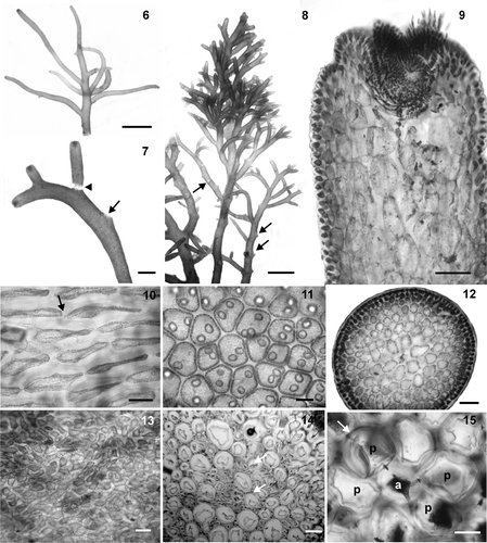
Figs 6–15. Branchlets and vegetative structures of Laurencia dendroidea. 6. Detail of long and sinuous ultimate branchlets. 7. Detail of a branch showing scar of released branchlet (arrow). Note detachment of a branchlet (arrowhead). 8. Upper portion of the thallus showing branches with many scars of released branchlets (arrows). 9. Longitudinal section through a branchlet showing projecting cortical cells. 10. Cortical cells in surface view, showing a secondary pit‐connection (arrow). 11. Cortical cells in surface view, showing corps en cerise in living material. 12. Transverse section of the thallus. 13. Old portion of the thallus in surface view showing secondary cortication. 14. Detail of medulla of older thallus in transverse section, showing filling cells (arrows). 15. Transverse section of the upper portion of a branch showing an axial cell (a) with four pericentral cells (p). Note lenticular thickening in pericentral cell (arrow). Scale bars = 2 mm (Figs , ), 500 µm (), 100 µm (Figs , , ), 50 µm (Figs , ) and 25 µm (Figs , ).
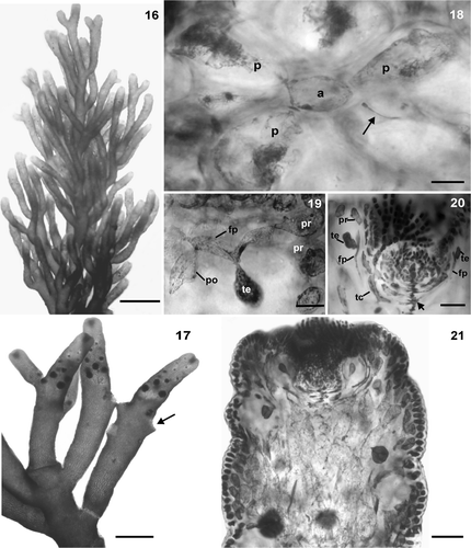
Figs 16–21. Tetrasporangial structures of Laurencia dendroidea. 16. Tetrasporangial branches. 17. Detail of tetrasporangial branchlets. Note scars of released branchlets (arrow). 18. Transverse section of tetrasporangial axial segment showing an axial cell (a) and one fertile pericentral cell, the fourth (arrow); the other pericentral cells remain vegetative (p). 19. Detail of a fertile pericentral cell (fp) with two pre‐sporangial cover cells (pr), tetrasporangium (te) and one post‐sporangial cover cell (po). 20. Longitudinal section through an apical portion of tetrasporangial branchlet, showing origin of tetrasporangia (te) from axial cell (arrow), fertile pericentral cells (fp), and pre‐sporangial cover cells (pr). Note trichoblast (tc) from an axial cell. 21. Longitudinal section through a tetrasporangial branchlet showing parallel arrangement of the tetrasporangia. Scale bars = 2 mm (), 500 µm (), 100 µm (), 50 µm (Figs , ), and 25 µm ().
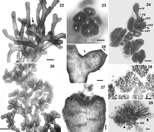
Figs 22–29. Female and male structures of Laurencia dendroidea. 22. Female branches. Note cystocarp (arrow). 23. Procarp‐bearing segment with five pericentral cells, the fifth becoming the supporting cell (su) of the carpogonial branch; central cell of procarp‐bearing segment (c). 24. Procarp before fertilization with four‐celled carpogonial branch (cb), carpogonium (cg), trichogyne (tr), lateral sterile group (ls), basal sterile group (bs), supporting cell (su), central cell of procarp‐bearing segment (c), basal cell of trichoblast (bt). 25. Longitudinal section through a female branchlet with prominent cystocarp without protuberant ostiole. 26. Male branches. 27. Longitudinal section through a male branchlet showing spermatangial branches in cup‐shaped tips. 28. Detail of trichoblast‐type spermatangial branches with terminal vesicular sterile cells. 29. Detail of spermatangial branches on trichoblast with two laterals, sterile (arrow) and spermatangial (arrowhead) branches on its suprabasal cell (sbt). Note spermatangia with an apical nucleus. Scale bars = 2 mm (), 1 mm (), 200 µm (), 100 µm (), 25 µm (Figs , ), 10 µm () and 5 µm ().
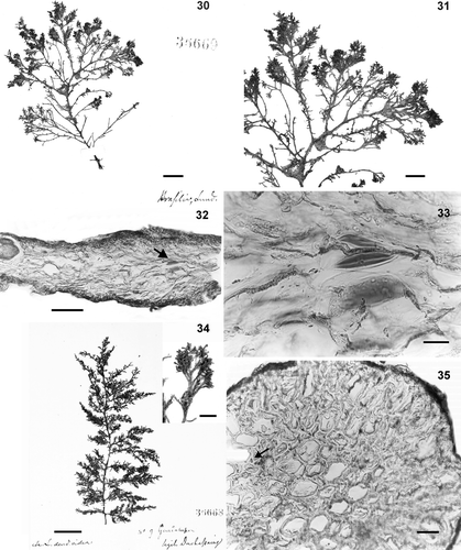
Figs 30–35. Type material of Laurencia dendroidea. . Lectotype (LD 36669). 30. Habit of the thallus. 31. Detail of branches. 32. Transverse section of ultimate branchlet showing lenticular thickenings (arrow). 33. Detail of lenticular thickenings. Figs , . Isolectotype (LD 36668). 34. Habit of the thallus. 35. Transverse section of old portion of the thallus showing filling cells (arrow). Scale bars = 2 cm (), 2 mm (, inset). 1 cm (), 5 mm (), 100 µm (), 40 µm () and 25 µm ().