Figures & data
Table 1. Optimized binary gradient system for the separation of phytoplankton lipophilic pigments on Waters Symmetry C18 column. Eluent A was 45 : 45 : 10 methanol : acetonitril : 1 M ammonium acetate (v/v/v) +3% 0.1 M ammonium acetate (v/v). Eluent B was 45 : 55 acetonitril : acetone (v/v).
Fig. 1. Mean content of the major lipophilic pigments chlorophyll a (Chl a), 4-hydroxymyxol glycoside (4-OH-Myxol), myxol glycoside (Myxol), echinenone (Echi) and β-carotene (β-Car) per dry weight (DW) (mg g−1) and carotenoid to chlorophyll a ratios (mg mg−1) of three temperate (hatched bars) and three tropical C. raciborskii strains (grey bars). Strains were grown at different combination of temperature (°C) and light intensity (µmol photons m−2 s−1) displayed as temperature/light intensity. Pigment content is given as mean and standard deviation of one replicate of three strains each (n = 3). Asterisks mark significant differences between temperate and tropical strains.
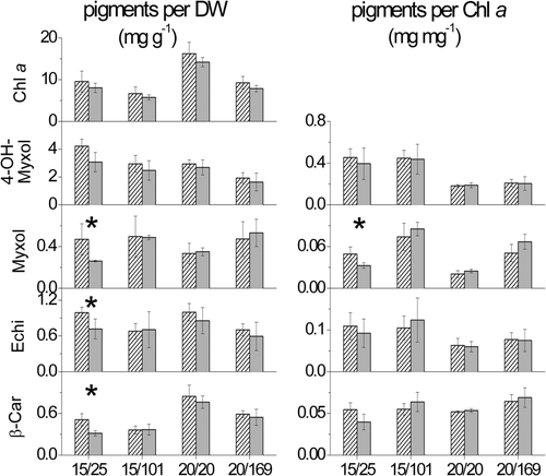
Fig. 2. Content of major lipophilic pigments per dry weight (DW) (mg g−1) and ratios of carotenoids (Car) to chlorophyll a (Chl a) (mg mg−1) of C. raciborskii strain 24G7 (A, C) and A. gracile strain 16D11 (B, D) grown at 80 µmol photons m−2 s−1 and seven temperatures from 10 to 35°C. Lines represent temperature-dependent growth curves (day−1) (A, B).
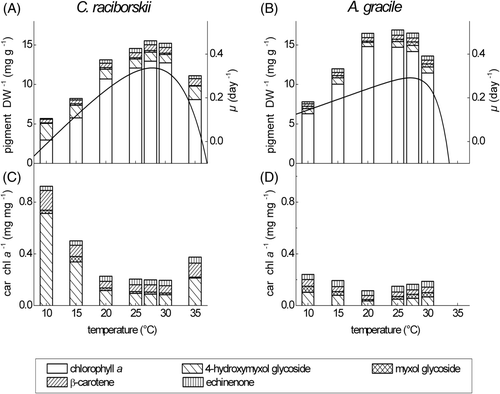
Table 2. Content of major lipophilic pigments per dry weight (DW) (mg g−1) and growth rates (day−1) of C. raciborskii strain 24G7 (C) and A. gracile strain 16D11 (A) acclimated to different temperatures and 80 µmol photons m−2 s−1. Mean and standard deviation of three pigment samples taken at different time-points during steady-state growth are given (n = 3). Differences of the pigment contents between the two species were tested for significance with t-test for independent samples and Mann–Whitney U-test. Abbreviations: ns non significant; *P ≤ 0.05; ** P ≤ 0.01; *** P ≤ 0.001.
Fig. 3. Mean content of the major pigments per dry weight (DW) (mg g−1) and light-dependent growth curves (day−1) of three German C. raciborskii strains (left) and three German A. gracile strains (right) grown at 20 and 15°C and four different light intensities. The growth curve is given as a fit of growth rates of the three strains each.
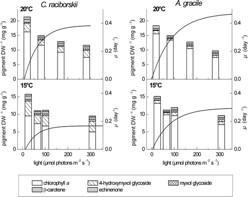
Fig. 4. Ratios of total carotenoids (Total Car), 4-hydroxymyxol glycoside (4-OH-Myxol), myxol glycoside (Myxol), echinenone (Echi) and β-carotene (β-Car) to chlorophyll a (Chl a), given as mean and standard deviation of three German C. raciborskii strains (black boxes) and three German A. gracile strains (open boxes). Cultures were grown at 20°C (left panels) and 15°C (right panels) and four different light intensities. Linear regression was performed without the carotenoid content at the lowest light intensity. The slope b of the regression curve and the coefficient of determination R 2 are displayed in standard font for C. raciborskii and in italics for A. gracile.
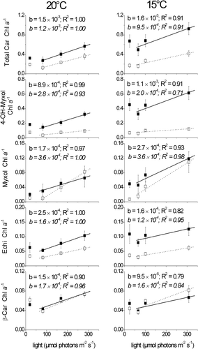
Table 3. Content of major lipophilic pigments per dry weight (DW) (mg g−1) and growth rates (day−1) of three German C. raciborskii (C) and three German A. gracile (A) strains acclimated to 15 and 20°C and different light intensities. Pigment content is given as the mean and standard deviation of one sample for each of three strains of each species (n = 3). Differences of the pigment contents between the two species were tested for significance with t-test for independent samples and Mann–Whitney U-test. Abbreviations: ns non significant; *P ≤ 0.05; ** P ≤ 0.01; *** P ≤ 0.001.
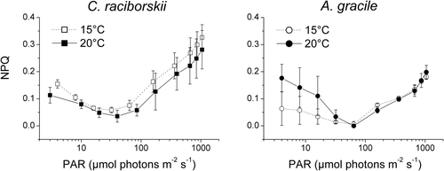
Fig. 5. Non-photochemical quenching of fluorescence (NPQ) plotted against photosynthetically active radiation (PAR) emitted by the PHYTO-PAM device. Three C. raciborskii (left panel) and three A. gracile (right panel) strains were grown at 88 µmol photons m−2 s−1 and 15°C (open symbols) and 20°C (closed symbols), respectively. NPQ is given as mean and standard deviation of the measurements of three different strains per species.