Figures & data
Fig. 1. The study areas and the locations of sampling sites.
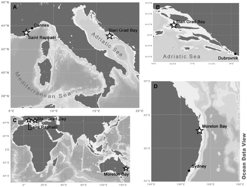
Table 1. List of samples from the Adriatic Sea checked for the presence of Cocconeis caulerpacola. Occurrence of C. caulerpacola estimated as + to +++ (+ present in moderate quantities, ++ abundant, +++ extremely abundant) or T (trace); – indicates that the species was not observed in the sample.
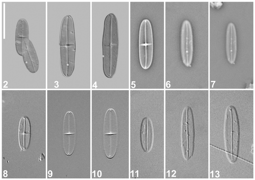
Figs 2–13. Cocconeis caulerpacola, LM. Specimens from the holotype slide. are micrographs taken by means of an advanced photomicrography method (kindly provided by Wulf Herwig); are phase contrast; are differential interference contrast. 2. Holotype specimen, comprising raphe (left) and sternum (right) valves. 3–13. Selected raphe (, ) and sternum () valves. Scale bar = 10 µm.
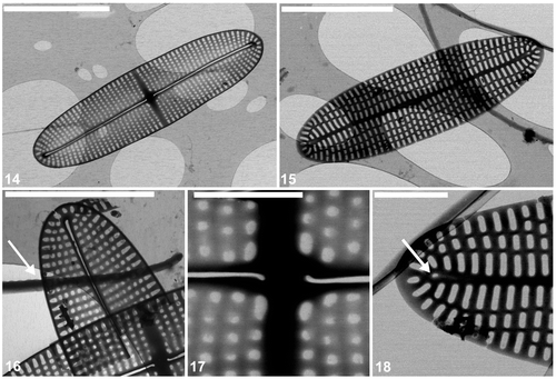
Figs 14–18. Cocconeis caulerpacola, TEM. 14. Raphe valve showing the striation and raphe branches with terminal and central endings. Note that the raphe endings are slightly bent towards opposite sides. 15. Sternum valve with longitudinal ribs and transapically elongate areolae. 16. Detail of raphe valve apex and part of valvocopula with very shallow undulations on one margin (arrow). 17. Centre of raphe valve. 18. Apex of sternum valve. Note the rudiment of a raphe (arrow). Scale bars = 5 µm (), 4 µm (); and 1 µm ().
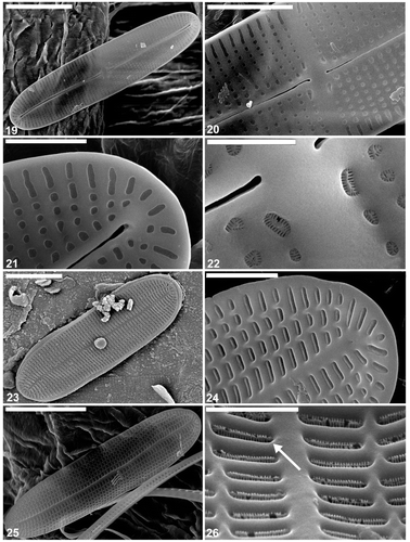
Figs 19–26. Cocconeis caulerpacola, SEM, from the holotype sample, Croatia. 19, 20. Raphe valve exterior, general view. Note sigmoid raphe with external central and terminal endings slightly expanded. 21. Internal view of raphe valve apex. Note the terminal raphe ending. 22. Close up of the centre of the interior of the raphe valve. 23. Sternum valve, external view. 24. Internal view of the sternum valve apex. 25. Sternum valve interior. 26. Close up of sternum valve interior. Note the areolae occlusions (arrow) containing apically oriented slits. Scale bars = 5 µm (); 3 µm (); 2 µm (); 1 µm (); and 500 nm ().
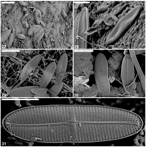
Figs 27–31. Cocconeis caulerpacola, SEM, from France. 27, 28. Specimens photographed in situ on Caulerpa taxifolia. 29, 30. External and internal views of cleaned sample. 31. Raphe valves, internal view. Note the internal central and apical raphe endings. Scale bars = 10 µm (); 6 µm (); 4 µm (); and 3 µm (,).
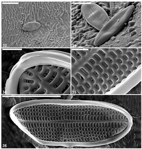
Figs 32–36. Cocconeis caulerpacola from France, SEM. show cells in situ on Caulerpa taxifolia. 32, 33. Cells with sternum valve uppermost, showing generally convex surface along the margins then becoming flat with a slightly depressed sternum. Note that the transapical striae are parallel in the middle, becoming radiate towards the apices. 34–36. Close up and whole () of sternum valve interior showing transapically elongate areolae. The areolae contain the same type of hymenate occlusions as in the raphe valve. Scale bars = 6 µm (); 4 µm (); 3 µm (); and 500 nm ().
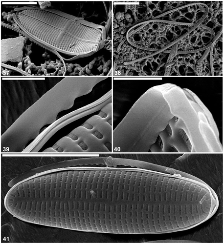
Figs 37–41. Cocconeis caulerpacola from France, SEM. 37. Frustule in oblique view. 38. Valvocopulae detached from frustules of C. caulerpacola. 39, 40. Close ups of valvocopulae: the pars interior has an undulate margin fitting over the valve interstriae. 41. Whole frustule with detached valvocopula. Scale bars = 4 µm (); 3 µm (); and 500 nm ().
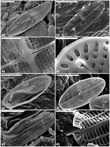
Figs 42–49. Cocconeis caulerpacola from Australia, SEM. 42. Raphe valve, external view. Note the sigmoid raphe and the areola pattern typical for C. caulerpacola from the type habitat. 43–45. Raphe valve internal view. Note the hymenate areolae occlusions. 46, 47. Sternum valve external view. 48, 49. Sternum valve internal view. Note the structure of the areolae hymenate occlusions. Scale bars = 5 µm (); 4 µm (); 3 µm (); 1 µm (); and 500 nm ().