Figures & data
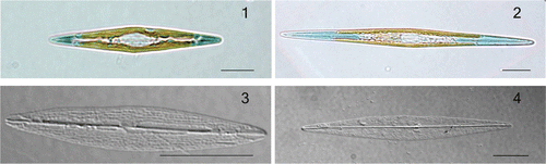
Figs 1–4. Haslea karadagensis and H. ostrearia, LM. 1. Live cell of H. karadagensis. 2. Live cell of H. ostrearia. 3. Cleaned frustule of H. karadagensis. 4. Cleaned frustule of H. ostrearia. Scale bars = 10 µm.
Figs 5–9. Haslea karadagensis, SEM. 5. External view of an entire valve. 6. External view of valve centre, showing the longitudinal slits and the straight proximal raphe fissures. 7. Internal view of valve centre, showing the rectangular areolae and the straight proximal raphe ends. 8. External view of apex showing the ± straight distal ending, longitudinal slits, and the continuous peripheral slit. 9. Internal view of apex showing the straight helictoglossa and the rectangular areolae. Scale bars = 10 µm () or 1 µm ().
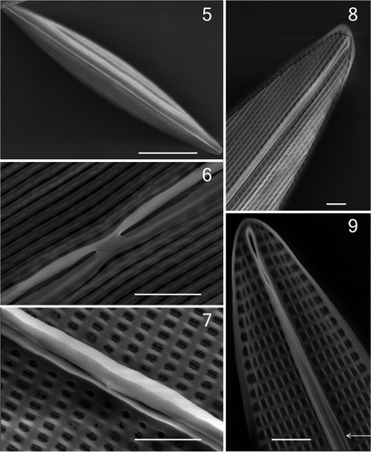
Table 1. Morphological data on Haslea karadagensis and H. ostrearia (this study), compared with those on H. ostrearia (or former Navicula ostrearia) and H. pseudostrearia obtained from the literature. When available from the literature, observed extremes are in bold. n.d. = no data.
Figs 10, 11. Distribution of lengths and widths in Haslea karadagensis and H. ostrearia. 10. Values measured in natural populations of H. karadagensis collected between April and May 2008 (n = 1148 cells). 11. Relationship between mean values of width and length measured on the different clones of both species maintained in cultures. The equations of the fitting lines are: y = 0.01 x + 8.21 for H. karadagensis (n = 22, R2 = 0.08), y = 0.03 x + 4.28 for H. ostrearia (n = 21, R2 = 0.63). Also included are mean values from available literature for Haslea/Navicula ostrearia from (a) Molisch (Citation1903), (b) Proshkina-Lavrenko (Citation1963), (c) Robert (Citation1973), (d) Massé et al. (Citation2001).
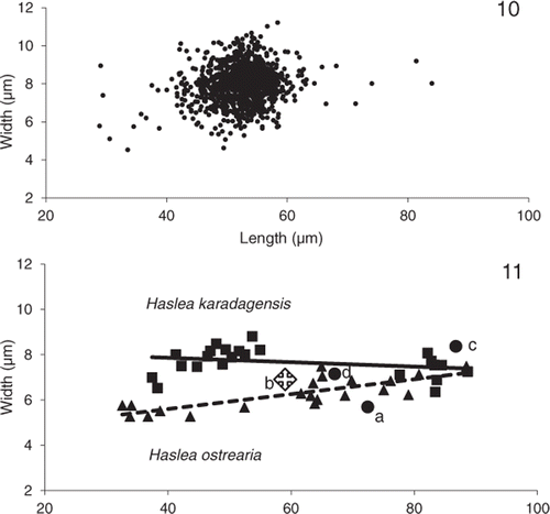
Fig. 12. Absorbance spectra, from 200 to 800 nm, of the pigments released into the medium by H. ostrearia and H. karadagensis. Inset: detail of the absorbance spectra from 550 to 750 nm, with expanded vertical scale.
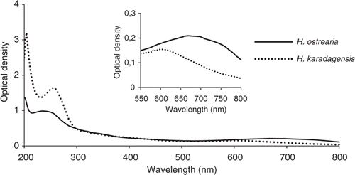
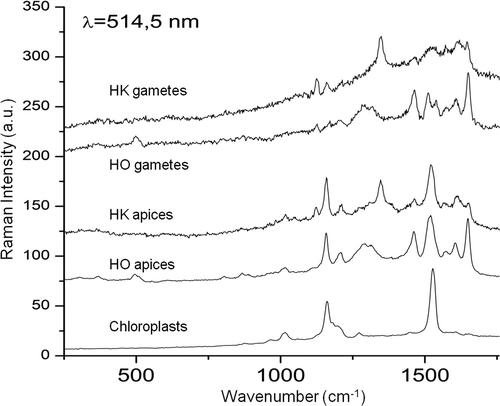
Fig. 13. Raman spectra at 514.5 nm, recorded using vegetative cells in exponential growth phase or gametes from H. karadagensis (HK) and H. ostrearia (HO). The laser was pointed at the coloured areas (apices of vegetative cells, gametes), or at the chloroplasts (vegetative cells).