1,351
Views
16
CrossRef citations to date
0
Altmetric
Original Articles

Taxonomic reassessment of Polysiphonia foetidissima (Rhodomelaceae, Rhodophyta) and similar species, including P. schneideri, a newly introduced species in Europe

, , , &
Pages 345-362 | Received 11 Oct 2012, Accepted 30 Apr 2013, Published online: 10 Oct 2013

Figures & data

Fig. 1. Maximum likelihood tree for Polysiphonia and their relatives from plastid-encoded rbcL sequence data. The bootstrap values shown above the branches are from 1000 bootstrap resamplings.
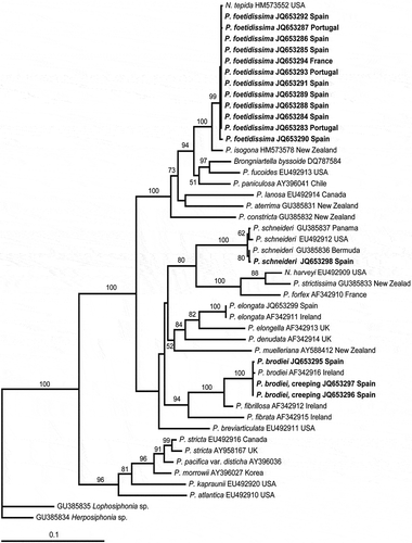
Fig. 1. Maximum likelihood tree for Polysiphonia and their relatives from plastid-encoded rbcL sequence data. The bootstrap values shown above the branches are from 1000 bootstrap resamplings.

Figs 2–6. Polysiphonia foetidissima: type material. 2. Lectotype (PC 0146017). 3. Cross section of an axis with 8 pericentral cells. 4. Rhizoids cut off from pericentral cells (arrow). 5. Apex showing a branch formed in the axil of a trichoblast (arrow) and trichoblasts or scar cells several segments apart (arrowheads). 6. Axes with a scar cell of a trichoblast (arrowhead). Scale bars = 1.5 cm (), 25 µm () and 90 µm ().
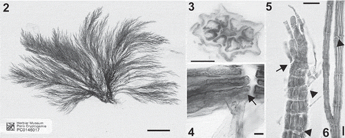
Figs 2–6. Polysiphonia foetidissima: type material. 2. Lectotype (PC 0146017). 3. Cross section of an axis with 8 pericentral cells. 4. Rhizoids cut off from pericentral cells (arrow). 5. Apex showing a branch formed in the axil of a trichoblast (arrow) and trichoblasts or scar cells several segments apart (arrowheads). 6. Axes with a scar cell of a trichoblast (arrowhead). Scale bars = 1.5 cm (Fig. 2), 25 µm (Figs 3–5) and 90 µm (Fig. 6).

Figs 7–9. Polysiphonia foetidissima: vegetative morphology of Iberian specimens. 7. Habit of vegetative thallus. 8. Erect axis with trichoblasts three or four segments apart and branches seven or eight segments apart. 9. Creeping axis with rhizoids cut off from pericentral cells and two adventitious branches. Scale bars = 1 mm () and 100 µm ().
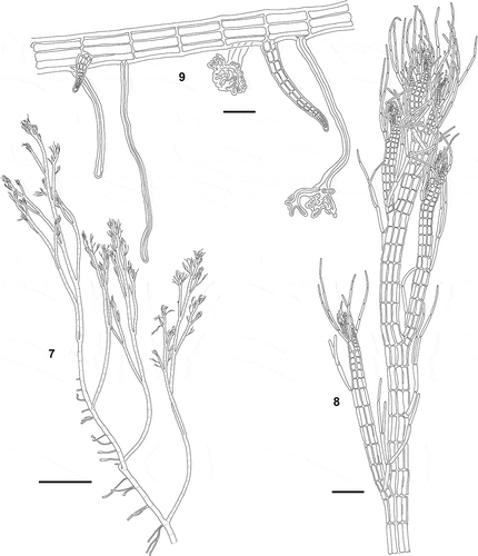
Figs 7–9. Polysiphonia foetidissima: vegetative morphology of Iberian specimens. 7. Habit of vegetative thallus. 8. Erect axis with trichoblasts three or four segments apart and branches seven or eight segments apart. 9. Creeping axis with rhizoids cut off from pericentral cells and two adventitious branches. Scale bars = 1 mm (Fig. 7) and 100 µm (Figs 8, 9).

Figs 10–17. Polysiphonia foetidissima: vegetative morphology of Iberian specimens. 10. Entangled decumbent axes on a turf of Rhodothamniella floridula. 11, 12. Erect axes with trichoblasts 4 segments apart and branches 7 or 8 segments apart. 13–17. Cross- section of axes with 6–9 pericentral cells. Scale bars = 1 cm (), 400 µm (), 60 µm () and 50 µm ().
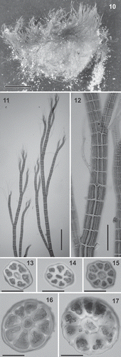
Figs 10–17. Polysiphonia foetidissima: vegetative morphology of Iberian specimens. 10. Entangled decumbent axes on a turf of Rhodothamniella floridula. 11, 12. Erect axes with trichoblasts 4 segments apart and branches 7 or 8 segments apart. 13–17. Cross- section of axes with 6–9 pericentral cells. Scale bars = 1 cm (Fig. 10), 400 µm (Fig. 11), 60 µm (Fig. 12) and 50 µm (Figs 13–17).

Figs 18–21. Polysiphonia foetidissima: reproductive morphology of Iberian specimens. 18. Branch with tetrasporangia in spiral series. 19. Mature procarp showing the axial cell (ax), the supporting cell (su), one-celled second sterile group (st2) and four-celled carpogonial branch (1–4). 20. Erect axis with cystocarps. 21. Apical portion of an erect axis with cylindrical spermatangial branches. Scale bars = 100 µm (), and 10 µm ().
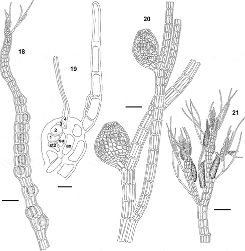
Figs 18–21. Polysiphonia foetidissima: reproductive morphology of Iberian specimens. 18. Branch with tetrasporangia in spiral series. 19. Mature procarp showing the axial cell (ax), the supporting cell (su), one-celled second sterile group (st2) and four-celled carpogonial branch (1–4). 20. Erect axis with cystocarps. 21. Apical portion of an erect axis with cylindrical spermatangial branches. Scale bars = 100 µm (Figs 18, 20, 21), and 10 µm (Fig. 19).

Figs 22–25. Polysiphonia foetidissima: reproductive morphology of Iberian specimens. 22. Branches with tetrasporangia in spiral series. 23. Erect axes with cystocarps. 24. Apical portions of erect axes with cylindrical spermatangial branches. 25. Forked spermatangial branch. Scale bars = 400 µm () and 50 µm ().
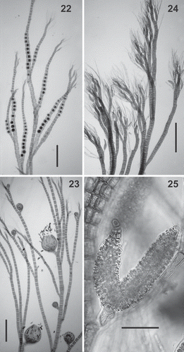
Figs 22–25. Polysiphonia foetidissima: reproductive morphology of Iberian specimens. 22. Branches with tetrasporangia in spiral series. 23. Erect axes with cystocarps. 24. Apical portions of erect axes with cylindrical spermatangial branches. 25. Forked spermatangial branch. Scale bars = 400 µm (Figs 22–24) and 50 µm (Fig. 25).

Fig. 26. Distribution of Polysiphonia foetidissima (circles) and P. schneideri (squares) along the Atlantic coasts of the Iberian Peninsula.
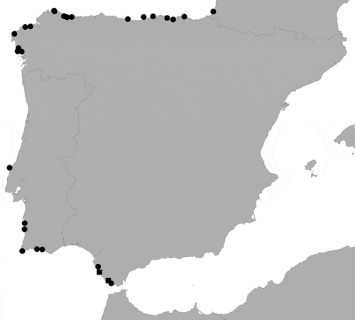
Fig. 26. Distribution of Polysiphonia foetidissima (circles) and P. schneideri (squares) along the Atlantic coasts of the Iberian Peninsula.

Figs 27–36. Polysiphonia schneideri: vegetative and reproductive morphology of Iberian specimens. 27. Habit of vegetative thallus. 28. Rhizoids cut off from pericentral cells (arrow). 29, 30. Cross-sections of axes with 6 or 7 pericentral cells. 31. Branches growing in the axils of trichoblasts (arrow). 32. Trichoblasts borne several segments apart (arrowheads). 33. Tetrasporangia in straight series. 34. Procarp with four-celled carpogonial branch (1–4). 35. Mature cystocarp. 36. Spermatangial branch formed at one of the branches of the first dichotomy of a trichoblast. Scale bars = 0.5 cm (), 100 µm (), 50 µm (), 200 µm () and 25 µm ().
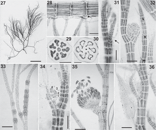
Figs 27–36. Polysiphonia schneideri: vegetative and reproductive morphology of Iberian specimens. 27. Habit of vegetative thallus. 28. Rhizoids cut off from pericentral cells (arrow). 29, 30. Cross-sections of axes with 6 or 7 pericentral cells. 31. Branches growing in the axils of trichoblasts (arrow). 32. Trichoblasts borne several segments apart (arrowheads). 33. Tetrasporangia in straight series. 34. Procarp with four-celled carpogonial branch (1–4). 35. Mature cystocarp. 36. Spermatangial branch formed at one of the branches of the first dichotomy of a trichoblast. Scale bars = 0.5 cm (Fig. 27), 100 µm (Fig. 28), 50 µm (Figs 29–32, 36), 200 µm (Figs 33, 35) and 25 µm (Fig. 34).

Fig. 37. World distribution map of Polysiphonia foetidissima and related species. P. foetidissima (● type locality, ● reports with descriptions, ○ reports), P. isogona (▬ type locality, ▬ reports with descriptions), P. kappannae (♦ type locality), P. nizamudinii (▼ type locality), P. schneideri (* type locality), P. stuposa (■ type locality, □ reports) and P. tepida (▲ type locality, ▲ reports with descriptions, ∆ reports). References: 1, Kützing (Citation1864); 2, Bornet (Citation1892); 3, Collins & Hervey (Citation1917); 4, Howe (Citation1918); 5, Batten (Citation1923); 6, Newton (Citation1931); 7, Hollenberg (Citation1958); 8, Lancelot (Citation1966); 9, Sreenivasa Rao (Citation1967); 10, Oliveira Filho (Citation1969); 11, Hollenberg (Citation1968); 12, Giaccone (Citation1969); 13, Ardré (Citation1970); 14, Cordeiro-Marino (Citation1977); 15, Kapraun (Citation1977); 16, Farooqui & Begum (Citation1978); 17, Giaccone (Citation1978); 18, Kapraun (Citation1979); 19, Taylor (Citation1960); 20, Schnetter & Schnetter (Citation1981); 21, Weisscher (Citation1983); 22, Audiffred & Prud´homme van Reine (Citation1985); 23, Giaccone et al. (Citation1985); 24, Conde & Soto (Citation1986); 25, Silva et al. (Citation1987); 26, Ballesteros (Citation1990); 27, Adams (Citation1991); 28, Schneider & Searles (Citation1991); 29, Ballesteros (Citation1993); 30, Lawson et al. (Citation1995); 31, Silva et al. (Citation1996); 32, Abbott (Citation1999); 33, Coll & Oliveira (Citation1999); 34, Rojas-González & Afonso-Carrillo (Citation2000, Citation2008); 35, McDermid et al. (Citation2002); 36, Womersley (Citation2003); 37, Guimarães et al. (Citation2004); 38, Suárez (Citation2005); 39, Duncan & Lee Lum (Citation2006); 40, Tsuda et al. (Citation2006); 41, Dawes & Mathieson (Citation2008); 42, Taskin et al. (Citation2008); 43, Stuercke & Freshwater (Citation2010); 44, Mamoozadeh & Freshwater (Citation2011).
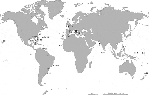
Fig. 37. World distribution map of Polysiphonia foetidissima and related species. P. foetidissima (● type locality, ● reports with descriptions, ○ reports), P. isogona (▬ type locality, ▬ reports with descriptions), P. kappannae (♦ type locality), P. nizamudinii (▼ type locality), P. schneideri (* type locality), P. stuposa (■ type locality, □ reports) and P. tepida (▲ type locality, ▲ reports with descriptions, ∆ reports). References: 1, Kützing (Citation1864); 2, Bornet (Citation1892); 3, Collins & Hervey (Citation1917); 4, Howe (Citation1918); 5, Batten (Citation1923); 6, Newton (Citation1931); 7, Hollenberg (Citation1958); 8, Lancelot (Citation1966); 9, Sreenivasa Rao (Citation1967); 10, Oliveira Filho (Citation1969); 11, Hollenberg (Citation1968); 12, Giaccone (Citation1969); 13, Ardré (Citation1970); 14, Cordeiro-Marino (Citation1977); 15, Kapraun (Citation1977); 16, Farooqui & Begum (Citation1978); 17, Giaccone (Citation1978); 18, Kapraun (Citation1979); 19, Taylor (Citation1960); 20, Schnetter & Schnetter (Citation1981); 21, Weisscher (Citation1983); 22, Audiffred & Prud´homme van Reine (Citation1985); 23, Giaccone et al. (Citation1985); 24, Conde & Soto (Citation1986); 25, Silva et al. (Citation1987); 26, Ballesteros (Citation1990); 27, Adams (Citation1991); 28, Schneider & Searles (Citation1991); 29, Ballesteros (Citation1993); 30, Lawson et al. (Citation1995); 31, Silva et al. (Citation1996); 32, Abbott (Citation1999); 33, Coll & Oliveira (Citation1999); 34, Rojas-González & Afonso-Carrillo (Citation2000, Citation2008); 35, McDermid et al. (Citation2002); 36, Womersley (Citation2003); 37, Guimarães et al. (Citation2004); 38, Suárez (Citation2005); 39, Duncan & Lee Lum (Citation2006); 40, Tsuda et al. (Citation2006); 41, Dawes & Mathieson (Citation2008); 42, Taskin et al. (Citation2008); 43, Stuercke & Freshwater (Citation2010); 44, Mamoozadeh & Freshwater (Citation2011).

Figs 38–42. Polysiphonia stuposa: type material. 38. Holotype (MEL 2324435, collected by Sandri in Dalmatia, Croatia). 39. Prostrate axis with rhizoids cut off from pericentral cells. 40. Apex of an erect axis showing branch origin not associated with trichoblasts. 41. Cross section of an axis with 7 pericentral cells. 42. Erect axes. Scale bars = 1 cm (), 150 µm (), 30 µm (), 20 µm () and 600 µm ().

Figs 38–42. Polysiphonia stuposa: type material. 38. Holotype (MEL 2324435, collected by Sandri in Dalmatia, Croatia). 39. Prostrate axis with rhizoids cut off from pericentral cells. 40. Apex of an erect axis showing branch origin not associated with trichoblasts. 41. Cross section of an axis with 7 pericentral cells. 42. Erect axes. Scale bars = 1 cm (Fig. 38), 150 µm (Fig. 39), 30 µm (Fig. 40), 20 µm (Fig. 41) and 600 µm (Fig. 42).

Figs 43–47. Polysiphonia tepida: type material. 43. Holotype (MICH, Herbarium of W.R. Taylor, collected by H.L. Blomquist in 1940 in Beaufort, North Carolina, U.S.A.). 44. Cross-section of an axis with eight pericentral cells. 45. Creeping axis with a rhizoid cut off from a pericentral cell. 46. Apical portions of an erect axis with abundant trichoblasts borne 3 or 4 segments apart and branches arising from the axils of trichoblasts. 47. Scar cell of a trichoblast on an erect axis. Scale bars = 2 cm () and 50 µm ().
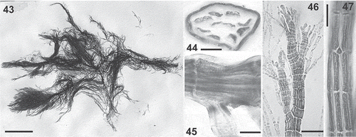
Figs 43–47. Polysiphonia tepida: type material. 43. Holotype (MICH, Herbarium of W.R. Taylor, collected by H.L. Blomquist in 1940 in Beaufort, North Carolina, U.S.A.). 44. Cross-section of an axis with eight pericentral cells. 45. Creeping axis with a rhizoid cut off from a pericentral cell. 46. Apical portions of an erect axis with abundant trichoblasts borne 3 or 4 segments apart and branches arising from the axils of trichoblasts. 47. Scar cell of a trichoblast on an erect axis. Scale bars = 2 cm (Fig. 43) and 50 µm (Figs 44–47).

Figs 48–55. Polysiphonia exilis: type material. 48. Holotype (TCD 0012730, collected by Harvey at Key West, Florida). 49. Erect axes with secund branches. 50. Cross-section of thallus with nine pericentral cells. 51. Rhizoid cut off from a pericentral cell (arrow). 52. Detail of an erect axis with scar cells of trichoblasts on every segment (arrows) and pericentral cells in straight rows. 53. Detail of an erect axis with young cicatrigenous branches growing from scar cells (arrowheads); the arrow shows a scar cell of a trichoblast. 54. Detail of an axis bearing a branch with young tetrasporangia forming a spiral series; the arrowhead indicates a young cicatrigenous branch. 55. Detail of a prostrate axis bearing erect axes at irregular intervals. Scale bars = 2.5 cm (), 3 mm (), 50 µm (), 250 µm () and 650 µm ().
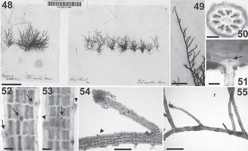
Figs 48–55. Polysiphonia exilis: type material. 48. Holotype (TCD 0012730, collected by Harvey at Key West, Florida). 49. Erect axes with secund branches. 50. Cross-section of thallus with nine pericentral cells. 51. Rhizoid cut off from a pericentral cell (arrow). 52. Detail of an erect axis with scar cells of trichoblasts on every segment (arrows) and pericentral cells in straight rows. 53. Detail of an erect axis with young cicatrigenous branches growing from scar cells (arrowheads); the arrow shows a scar cell of a trichoblast. 54. Detail of an axis bearing a branch with young tetrasporangia forming a spiral series; the arrowhead indicates a young cicatrigenous branch. 55. Detail of a prostrate axis bearing erect axes at irregular intervals. Scale bars = 2.5 cm (Fig. 48), 3 mm (Fig. 49), 50 µm (Figs 50–53), 250 µm (Fig. 54) and 650 µm (Fig. 55).

Figs 56–61. Polysiphonia brodiei: small and creeping forms in the Iberian Peninsula. 56. Habit of a tetrasporophyte growing on Codium adhaerens. 57. Spermatangial branches borne on every segment and without sterile apical cells. 58. Erect axes with scar cells of trichoblasts on every segment. 59–61. Upper portions of erect axes with branches arising in the axils of trichoblasts, 3 or 4 segments apart. Scale bars = 2 cm (), 80 µm (), 50 µm (), 500 µm () and 200 µm ().
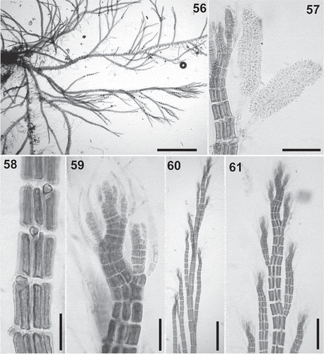
Figs 56–61. Polysiphonia brodiei: small and creeping forms in the Iberian Peninsula. 56. Habit of a tetrasporophyte growing on Codium adhaerens. 57. Spermatangial branches borne on every segment and without sterile apical cells. 58. Erect axes with scar cells of trichoblasts on every segment. 59–61. Upper portions of erect axes with branches arising in the axils of trichoblasts, 3 or 4 segments apart. Scale bars = 2 cm (Fig. 56), 80 µm (Fig. 57), 50 µm (Figs 58, 59), 500 µm (Fig. 60) and 200 µm (Fig. 61).

Table 1. Main taxonomic features of Polysiphonia foetidissima and similar species with 6–11 pericentral cells, rhizoids cut off from pericentral cells, and prostrate or creeping habit.

Table S1. Materials collected during this study, including herbarium codes and Genbank accession numbers.

Supplemental material

Supplementary material

Download MS Word (111 KB)

Reprints and Corporate Permissions

Please note: Selecting permissions does not provide access to the full text of the article, please see our help page How do I view content?

To request a reprint or corporate permissions for this article, please click on the relevant link below:

Academic Permissions

Please note: Selecting permissions does not provide access to the full text of the article, please see our help page How do I view content?

Obtain permissions instantly via Rightslink by clicking on the button below:

If you are unable to obtain permissions via Rightslink, please complete and submit this Permissions form. For more information, please visit our Permissions help page.