Figures & data
Table 1. Laurencia complex species reported from Lusitanian Macaronesia, with sources of information.
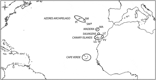
Fig. 1. Map of Atlantic Ocean showing the Macaronesian archipelagos. Sites where Macaronesian specimens were collected in the present study are in the Azores: Pico (PI: Pocinho-Monte Candelaria 38.49686466° N/ 28.53992863° W; Barca-Madalena 38.53988167° N/ 28.52058390° W; Prainha do Norte 38.47720139° N/ 28.20443794° W; Lajes do Pico-Poça de Baleia 38.38998195° N/ 28.25144459 W; Lajes do Pico-Fábrica de Baleia 38.38862491° N/ 29. 19430601° W; Santa Cruz Ribeiras 38.404° N/ 28.1872° W), São Miguel (SM: Cerco da Caloura-Baía 37.7570843° N/ 25.81716330° W; Ferraria 37.8579856° N/ 25.85265462° W; Mosteiros 37.8992112° N/ 25.82103420° W), Santa Maria (SMa: Boca de Ribeira Seca 36.94337327° N/ 25.16456403° W; Emissores 36.99720678° N/ 25.17678807° W; Anjos-Este 37.00542551° N/ 25.16444382° W; Anjos-Ponta dos Frades 37.00788999° N/ 25.15019092° W; Anjos-Piscinas 37.00458430° N/ 25.15727202° W); in Madeira (MA: Seixal-Praia da Laje 32.82554110° N/ 17.11529253° W; Porto Moniz-Piscinas 32.86802811° N/ 17.17135116° W; Ponta de Sao Jorge-Casi 32.8357° N/ 16.9053° W); in the Canary Islands: Fuerteventura (FV: Garcey 28.345492° N/ 14.178111° W; El Cotillo 28.7013° N/ 14.0182° W), Lanzarote (LZ: Arrecife 28.957972° N/ 13.544525° W; Pechigueras 28.855217° N/ 13.872631° W), La Gomera (LG: El Charco de las Condesas 28.0839772° N/ 17.33672469° W; El Charco del Conde 28.05150° N/ 17.20269° W; Punta de la Dama 28.031° N/ 17.183° W), Tenerife (TF: El Pris 28.50981317° N/ 16.42174616° W; Puerto de la Cruz 28.4175° N/ 165462° W; Punta del Hidalgo 28.5739° N/ 16.5462° W).
Figs 2–5. Laurencia pyramidalis: type specimens from the herbarium of the Naturalis Biodiversity Center (section NHN), Leiden (L). 2. Herbarium sheet L 0820668 bearing lectotypes and syntypes (Herbarium Kützing). 3. Syntype, enlarged from 4. Syntype, enlarged from 5. Lectotype, enlarged from
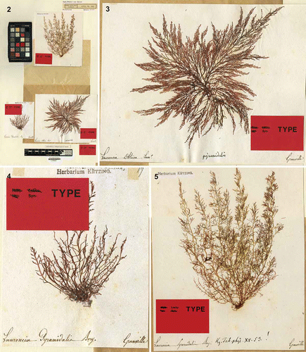
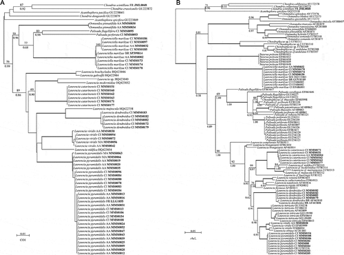
Fig. 6. Phylogram generated using neighbour-joining analyses from COI-5P sequences (A) and phylogenetic tree generated using Bayesian inference inferred from rbcL sequences (B). Voucher numbers (in bold) and the geographical origin of each specimen for which sequences were generated in the current study are indicated. FR: France, AA: Azores archipelago, MA: Madeira, CI: Canary Islands, MX: Mexico, BR: Brazil. Bootstrap values > 60% are indicated above nodes and Bayesian posterior probabilities are indicated under nodes. Bold branches indicate strongly supported nodes (bootstrap values > 95% and Bayesian probability > 0.99).
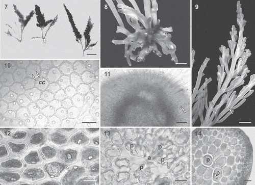
Figs 7–14. Laurencia pyramidalis from the Lusitanian Macaronesian region. 7. Habit. The main axes have sparse branching in the lower portions and first order laterals that decrease in length upwards and bear three further orders of branching in the same arrangement, resulting in a thallus that is pyramidal in outline. Scale bar = 2 cm. 8. Basal anchorage crust with stoloniferous branches. Scale bar = 1 mm. 9. Spiral branching with three or four orders of whorled branches. Branches issued from the first-order branches curved markedly towards the main axis, becoming almost parallel to it. Scale bar = 2 mm. 10. In surface view, cortical cells contain one or two corps en cerise (cc) in living specimens (arrows). Scale bar = 30 µm. 11. Branches with truncate tips showing dense hyaline trichoblasts with one corps en cerise per cell in living specimens (e.g. arrow). Scale bar = 50 µm. 12. Surface view showing polygonal cortical cells connected to each other by secondary pit connections (arrows). Scale bar = 30 µm. 13. Transverse section of the upper portion of a branch showing an axial cell (a) with four pericentral cells (p). Scale bar = 30 µm. 14. Transverse section of a thallus showing medullary cells with thickened walls. Note pericentral cells (p) with annular thickenings (arrow). Scale bar = 50 µm.
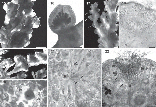
Figs 15–22. Laurencia pyramidalis from the Lusitanian Macaronesian region: reproductive structures. 15. Female gametophyte with cystocarps (arrows) located on the penultimate branches; they are subapical, sessile and prominent. Scale bar = 1 mm. 16. Longitudinal section showing a slightly pyriform cystocarp with a non-protuberant ostiole and clavate carposporangia. Scale bar = 200 µm. 17. Male gametophyte with spermatangial receptacles (arrows) located on the ultimate fertile branchlets. Scale bar = 500 µm. 18. Longitudinal section of a cup-shaped spermatangial receptacle. An axial cell (a) is discernible at the base bearing a fertile branch with many ovoid spermatangia. Scale bar = 50 µm. 19. Tetrasporangial plants with cylindrical branchlets. Scale bar = 2 mm. 20. Surface view of a tetrasporangium (te) with two presporangial cover cells (pr). Scale bar = 30 µm. 21. Transverse section near the apex of axial tetrasporangial segments with an axial cell (a), two vegetative pericentral cells (p1, p2) and two fertile pericentral cells (arrows). Scale bar = 30 µm. 22. Longitudinal section through a tetrasporangial branchlet showing the parallel arrangement of the tetrasporangia. Each fertile pericentral cell (arrows) cuts off presporangial cover cells (pr) distal to the initial tetrasporangium (te). Scale bar = 50 µm.
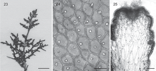
Figs 23–25. Laurenciella marilzae from the Azores. 23. Habit. Scale bar = 5 mm. 24. In surface view, cortical cells contain one corps en cerise (cc) in living specimens. Scale bar = 30 µm. 25. Longitudinal section through a branch showing corps en cerise in cortical and medullary cells. Scale bar = 10 µm.