1,492
Views
13
CrossRef citations to date
0
Altmetric
Original Articles

Systematics of green algae resembling Ulva conglobata, with a description of Ulva adhaerens sp. nov. (Ulvales, Ulvophyceae)

&
Pages 100-111 | Received 22 Dec 2013, Accepted 16 Aug 2014, Published online: 23 Jan 2015

Figures & data

Fig. 1. ML tree of Ulva based on rbcL sequences. Bootstrap values are indicated at branches (ML/MP/NJ). A bootstrap value of 100% is represented by *, while that below 50% is represented by -. Samples sequenced in this study are shown in bold. 1Blomster et al. (Citation1999). 2Tan et al. (Citation1999). 3Hayden et al. (Citation2003). 4Shimada et al. (Citation2003). 5Hiraoka et al. (Citation2004a). 6Hayden & Waaland (Citation2004). 7Loughnane et al. (Citation2008). 8Heesch et al. (Citation2009). 9Ichihara et al. (Citation2009). 10O’Kelly et al. (Citation2010). 11Kraft et al. (Citation2010). 12Mareš et al. (Citation2011).13Horimoto et al. (Citation2011). 14Ichihara et al. (Citation2013).
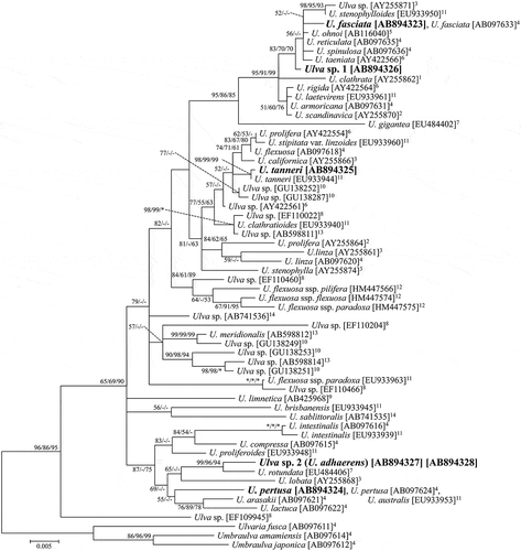
Fig. 1. ML tree of Ulva based on rbcL sequences. Bootstrap values are indicated at branches (ML/MP/NJ). A bootstrap value of 100% is represented by *, while that below 50% is represented by -. Samples sequenced in this study are shown in bold. 1Blomster et al. (Citation1999). 2Tan et al. (Citation1999). 3Hayden et al. (Citation2003). 4Shimada et al. (Citation2003). 5Hiraoka et al. (Citation2004a). 6Hayden & Waaland (Citation2004). 7Loughnane et al. (Citation2008). 8Heesch et al. (Citation2009). 9Ichihara et al. (Citation2009). 10O’Kelly et al. (Citation2010). 11Kraft et al. (Citation2010). 12Mareš et al. (Citation2011).13Horimoto et al. (Citation2011). 14Ichihara et al. (Citation2013).

Fig. 2. ML tree of Ulva based on ITS1 and ITS2 sequences. Bootstrap values are indicated at branches (ML/MP/NJ). A bootstrap value of 100% is represented by *, while that below 50% is represented by -. Samples sequenced in this study are shown in bold. 1Coat et al. (Citation1998). 2Blomster et al. (Citation1999). 3Tan et al. (Citation1999). 4Hayden et al. (Citation2003). 5Shimada et al. (Citation2003). 6Hiraoka et al. (Citation2004a). 7Hayden & Waaland (Citation2004). 8Kraft et al. (Citation2010). 9Mareš et al. (Citation2011).
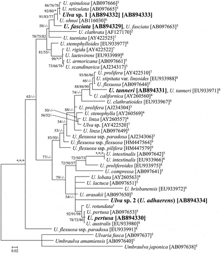
Fig. 2. ML tree of Ulva based on ITS1 and ITS2 sequences. Bootstrap values are indicated at branches (ML/MP/NJ). A bootstrap value of 100% is represented by *, while that below 50% is represented by -. Samples sequenced in this study are shown in bold. 1Coat et al. (Citation1998). 2Blomster et al. (Citation1999). 3Tan et al. (Citation1999). 4Hayden et al. (Citation2003). 5Shimada et al. (Citation2003). 6Hiraoka et al. (Citation2004a). 7Hayden & Waaland (Citation2004). 8Kraft et al. (Citation2010). 9Mareš et al. (Citation2011).

Figs 3–7. Representative fresh materials of green algae resembling Ulva conglobata. Fig. 3. U. fasciata. Fig. 4. U. pertusa. Fig. 5. U. tanneri. Fig. 6. Ulva sp. 1. Fig. 7. Ulva sp. 2 (U. adhaerens; fresh condition of the specimen designated for the holotype). Scale bars: 1 cm.
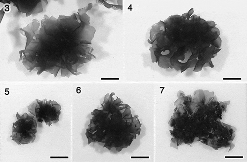
Figs 3–7. Representative fresh materials of green algae resembling Ulva conglobata. Fig. 3. U. fasciata. Fig. 4. U. pertusa. Fig. 5. U. tanneri. Fig. 6. Ulva sp. 1. Fig. 7. Ulva sp. 2 (U. adhaerens; fresh condition of the specimen designated for the holotype). Scale bars: 1 cm.

Figs 8–12. Morphology of Ulva sp. 1. Fig. 8. Marginal denticulations. Fig. 9. Surface view of the middle region. Fig. 10. Section near the base. Fig. 11. Section of the middle region. Fig. 12. Section near the margin. Scale bars: 50 μm.
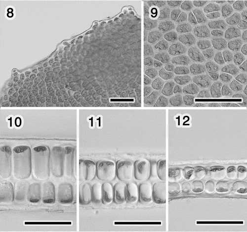
Figs 8–12. Morphology of Ulva sp. 1. Fig. 8. Marginal denticulations. Fig. 9. Surface view of the middle region. Fig. 10. Section near the base. Fig. 11. Section of the middle region. Fig. 12. Section near the margin. Scale bars: 50 μm.

Table 1. Summary of morphological characters of the specimens resembling U. conglobata, and of the original material of U. conglobata.

Figs 13–19. Morphology of Ulva sp. 2 (U. adhaerens). Fig. 13. Close-up of thallus, showing rhizoids in a distromatic region (arrowheads). Fig. 14. Surface view of the middle region. Fig. 15. Section near the rhizoid. Fig. 16. Section of the middle region. Fig. 17. Section near the margin. Fig. 18. Section of the lobes adhering with the rhizoids. Fig. 19. Close-up of the rhizoid. Scale bars: Fig. 13, 1 mm; Figs 14–19, 50 μm.
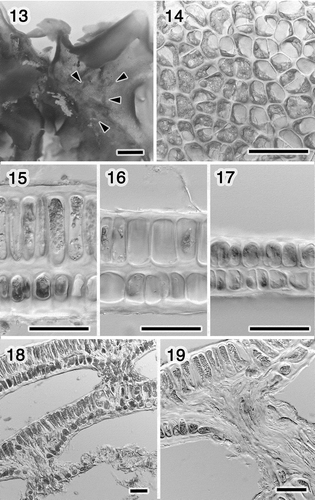
Figs 13–19. Morphology of Ulva sp. 2 (U. adhaerens). Fig. 13. Close-up of thallus, showing rhizoids in a distromatic region (arrowheads). Fig. 14. Surface view of the middle region. Fig. 15. Section near the rhizoid. Fig. 16. Section of the middle region. Fig. 17. Section near the margin. Fig. 18. Section of the lobes adhering with the rhizoids. Fig. 19. Close-up of the rhizoid. Scale bars: Fig. 13, 1 mm; Figs 14–19, 50 μm.

Figs 20–31. Ulva sp. 1. Swarmers and cultures grown at 20°C with a 14: 10 h light/dark cycle. Fig. 20. Swarmers. Fig. 21. Settled swarmer. Figs 22–26. Germling. Fig. 22. 4 days. Fig. 23. 6 days. Fig. 24. 10 days. Fig. 25. Two weeks. Fig. 26. Five weeks. Fig. 27. Thalli, 52 days. Fig. 28. Margin with an overlapped portion (arrowhead), 58 days. Fig. 29. Section of the overlapped marginal portion of the thallus shown in Fig. 28. Figs 30, 31. Thallus with holes due to releasing swarmers, 44 and 45 days. Scale bars: Figs 20–24, 10 μm; Figs 25, 26, 30, 31, 5 mm; Fig. 27, 1 cm; Fig. 28, 1 mm; Fig. 29, 50 μm.
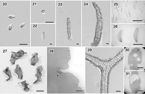
Figs 20–31. Ulva sp. 1. Swarmers and cultures grown at 20°C with a 14: 10 h light/dark cycle. Fig. 20. Swarmers. Fig. 21. Settled swarmer. Figs 22–26. Germling. Fig. 22. 4 days. Fig. 23. 6 days. Fig. 24. 10 days. Fig. 25. Two weeks. Fig. 26. Five weeks. Fig. 27. Thalli, 52 days. Fig. 28. Margin with an overlapped portion (arrowhead), 58 days. Fig. 29. Section of the overlapped marginal portion of the thallus shown in Fig. 28. Figs 30, 31. Thallus with holes due to releasing swarmers, 44 and 45 days. Scale bars: Figs 20–24, 10 μm; Figs 25, 26, 30, 31, 5 mm; Fig. 27, 1 cm; Fig. 28, 1 mm; Fig. 29, 50 μm.

Figs 32–48. Ulva sp. 2 (U. adhaerens). Swarmers and cultures grown at 15°C with a 12: 12 h light/dark cycle. Fig. 32. Swarmers. Fig. 33. Settled swarmer. Figs 34–38. Germling. Fig. 34. 5 days. Fig. 35. 6 days. Fig. 36. 9 days. Fig. 37. 20 days. Fig. 38. Five weeks. Fig 39. Thalli, 53 days. Fig. 40. Thalli, 62 days. Fig. 41. Margin with an overlapped portion (arrowhead), 49 days. Fig. 42. Section of the overlapped marginal portion of the thallus shown in Fig. 41. Fig. 43. Thallus having released swarmers from marginal portion, 62 days. The adhered regions with rhizoids look paler; arrowheads. Fig. 44. Thalli adhering near the bases (arrowheads), between the different surfaces (upper) or the same surfaces (lower). Fig. 45. Basal portion with the basipetal rhizoidal extensions, 31 days. Fig. 46. Longitudinal section of the connecting region of the distromatic lobes, 47 days. Fig. 47. Close-up of Fig. 46, showing rhizoidal extensions. Fig. 48. Section of the adhering region of the thallus shown in Fig. 44 (upper), showing secondary rhizoids. Scale bars: Figs 32–36, 10 μm; Figs 37, 38, 5 mm; Figs 39, 40, 43, 44, 1cm; Fig. 4, 1 mm; Figs 42, 45, 47, 48, 50 μm; Fig. 46, 200 μm.
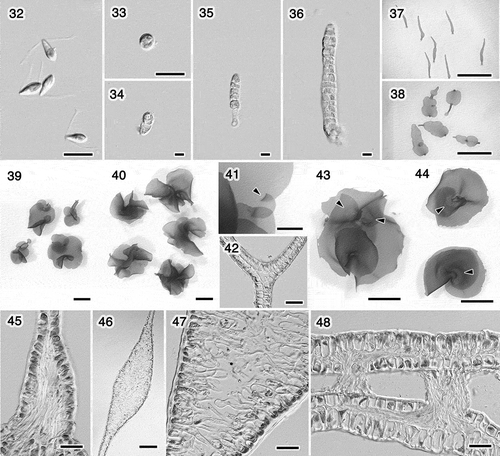
Figs 32–48. Ulva sp. 2 (U. adhaerens). Swarmers and cultures grown at 15°C with a 12: 12 h light/dark cycle. Fig. 32. Swarmers. Fig. 33. Settled swarmer. Figs 34–38. Germling. Fig. 34. 5 days. Fig. 35. 6 days. Fig. 36. 9 days. Fig. 37. 20 days. Fig. 38. Five weeks. Fig 39. Thalli, 53 days. Fig. 40. Thalli, 62 days. Fig. 41. Margin with an overlapped portion (arrowhead), 49 days. Fig. 42. Section of the overlapped marginal portion of the thallus shown in Fig. 41. Fig. 43. Thallus having released swarmers from marginal portion, 62 days. The adhered regions with rhizoids look paler; arrowheads. Fig. 44. Thalli adhering near the bases (arrowheads), between the different surfaces (upper) or the same surfaces (lower). Fig. 45. Basal portion with the basipetal rhizoidal extensions, 31 days. Fig. 46. Longitudinal section of the connecting region of the distromatic lobes, 47 days. Fig. 47. Close-up of Fig. 46, showing rhizoidal extensions. Fig. 48. Section of the adhering region of the thallus shown in Fig. 44 (upper), showing secondary rhizoids. Scale bars: Figs 32–36, 10 μm; Figs 37, 38, 5 mm; Figs 39, 40, 43, 44, 1cm; Fig. 41, 1 mm; Figs 42, 45, 47, 48, 50 μm; Fig. 46, 200 μm.

Figs 49–59. Morphology of the original material of U. conglobata. Fig. 49. Specimens of U. conglobata f. typica (1–7) and U. conglobata f. densa (8–11). Figs 50–52. Specimen no. 1. Fig. 50. Marginal denticulations. Fig. 51. Surface view. Fig. 52. Section. Figs 53, 54. Specimen no. 2. Fig. 53. Surface view. Fig. 54. Section. Figs 55, 56. Specimen no. 3. Fig. 55. Surface view. Fig. 56. Section. Figs 57–59. Specimen no. 4. Fig. 57. Marginal denticulation. Fig. 58. Surface view. Fig. 59. Section. Scale bars: Fig. 49, cm; Figs 50–59, 50 μm.
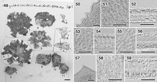
Figs 49–59. Morphology of the original material of U. conglobata. Fig. 49. Specimens of U. conglobata f. typica (1–7) and U. conglobata f. densa (8–11). Figs 50–52. Specimen no. 1. Fig. 50. Marginal denticulations. Fig. 51. Surface view. Fig. 52. Section. Figs 53, 54. Specimen no. 2. Fig. 53. Surface view. Fig. 54. Section. Figs 55, 56. Specimen no. 3. Fig. 55. Surface view. Fig. 56. Section. Figs 57–59. Specimen no. 4. Fig. 57. Marginal denticulation. Fig. 58. Surface view. Fig. 59. Section. Scale bars: Fig. 49, 1 cm; Figs 50–59, 50 μm.

Fig. 60. Ulva adhaerens. Photograph of the holotype herbarium specimen, TNS-AL 183435. Scale bar: 1 cm.
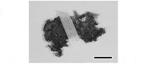
Fig. 60. Ulva adhaerens. Photograph of the holotype herbarium specimen, TNS-AL 183435. Scale bar: 1 cm.
Supplemental material

Supplementary Figs S1-S7

Download JPEG Image (981.4 KB)

Reprints and Corporate Permissions

Please note: Selecting permissions does not provide access to the full text of the article, please see our help page How do I view content?

To request a reprint or corporate permissions for this article, please click on the relevant link below:

Academic Permissions

Please note: Selecting permissions does not provide access to the full text of the article, please see our help page How do I view content?

Obtain permissions instantly via Rightslink by clicking on the button below:

If you are unable to obtain permissions via Rightslink, please complete and submit this Permissions form. For more information, please visit our Permissions help page.