996
Views
6
CrossRef citations to date
0
Altmetric
Original Articles

Morphology and molecular phylogeny of Thalassiosira sinica sp. nov. (Bacillariophyta) with delicate areolae and fultoportulae pattern

ORCID Icon, &
Pages 122-134 | Received 19 Jan 2017, Accepted 07 Aug 2017, Published online: 16 Jan 2018

Figures & data

Table 1. Sampling information for Thalassiosira strains.

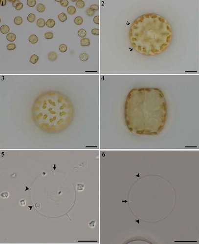
Figs 1–6. Thalassiosira sinica, LM. . Single cells. . Valve view showing organic threads (arrows) extending from valve margin. . Valve view indicating lobed chloroplasts. . Drum-shaped cell in girdle view. . Valve views of acidified specimens showing marginal fultoportulae (arrowheads) and rimoportulae (arrows). Scale bars: , 50 µm; , µm.

Figs 1–6. Thalassiosira sinica, LM. Fig. 1. Single cells. Fig. 2. Valve view showing organic threads (arrows) extending from valve margin. Fig. 3. Valve view indicating lobed chloroplasts. Fig. 4. Drum-shaped cell in girdle view. Figs 5, 6. Valve views of acidified specimens showing marginal fultoportulae (arrowheads) and rimoportulae (arrows). Scale bars: Fig. 1, 50 µm; Figs 2–6, 10 µm.

Figs 7, 8. Thalassiosira sinica, SEM. . External valve view. . Internal valve view. Scale bars: , , µm.

Figs 7, 8. Thalassiosira sinica, SEM. Fig. 7. External valve view. Fig. 8. Internal valve view. Scale bars: Figs 7, 8, 10 µm.

Figs 9–15. Thalassiosira sinica, SEM. . Central fultoportula without external development of the tube () and four or five satellite pores (, ) each possessing a pore cover (arrowheads in ) internally. . Rimoportula with longer and stronger tube on external valve. . Rimoportula, internal surface of the valve. . Detail of valves, showing marginal fultoportulae with longer tubes externally () than internally (), and valve-face fultoportulae lacking external tubes (), and a short internal tube is visible (). Scale bars: , 1 µm; , 2 µm.

Figs 9–15. Thalassiosira sinica, SEM. Figs 9–11. Central fultoportula without external development of the tube (Fig. 9) and four or five satellite pores (Figs 10, 11) each possessing a pore cover (arrowheads in Fig. 11) internally. Fig. 12. Rimoportula with longer and stronger tube on external valve. Fig. 13. Rimoportula, internal surface of the valve. Figs 14, 15. Detail of valves, showing marginal fultoportulae with longer tubes externally (Fig. 14) than internally (Fig. 15), and valve-face fultoportulae lacking external tubes (Fig. 14), and a short internal tube is visible (Fig. 15). Scale bars: Figs 9, 10, 12, 13, 1 µm; Fig. 11, 200 nm; Figs 14, 15, 2 µm.

Figs 16–19. Thalassiosira sinica, SEM. . Frustules in girdle views. . Enlargement of frustule part showing the band composition. eci, epicingulum; vc, valvocopula; c, copula; p, pleura, followed by number of pleurae. . Four bands of one valve. Scale bars: , 2 µm.

Figs 16–19. Thalassiosira sinica, SEM. Figs 16, 17. Frustules in girdle views. Fig. 18. Enlargement of frustule part showing the band composition. eci, epicingulum; vc, valvocopula; c, copula; p, pleura, followed by number of pleurae. Fig. 19. Four bands of one valve. Scale bars: Figs 16–19, 2 µm.

Fig. 20. Phylogenetic tree from Bayesian inference (BI) based on SSU rDNA sequences. Lampriscus kittonii makes up the outgroup. Posterior probability values are shown and asterisks indicate a value above 0.9. Thalassiosira sinica is shown in bold.

Fig. 20. Phylogenetic tree from Bayesian inference (BI) based on SSU rDNA sequences. Lampriscus kittonii makes up the outgroup. Posterior probability values are shown and asterisks indicate a value above 0.9. Thalassiosira sinica is shown in bold.

Fig. 21. Phylogenetic tree from Bayesian inference (BI) based on LSU sequences. Lampriscus kittonii makes up the outgroup. Posterior probabilities values are shown and asterisks indicate a value above 0.9. Thalassiosira sinica is shown in bold.

Fig. 21. Phylogenetic tree from Bayesian inference (BI) based on LSU sequences. Lampriscus kittonii makes up the outgroup. Posterior probabilities values are shown and asterisks indicate a value above 0.9. Thalassiosira sinica is shown in bold.

Fig. 22. Stylized drawings of the pattern of processes in Thalassiosira sinica and allied taxa. Fultoportulae are indicated by dots, rimoportulae by black ovals, and occluded processes by hollow ovals. Thalassiosira andamanica redrawn from Gedde (Citation1999), T. subtilis and T. diporocyclus from Hasle (Citation1972), T. sundarbana from Samanta & Bhadury. (Citation2015), T. tubifera and T. lundiana from Fryxell (Citation1975), T. thailandica from Boonyapiwat & Takano (Citation1986), T. sinica from this study.

Fig. 22. Stylized drawings of the pattern of processes in Thalassiosira sinica and allied taxa. Fultoportulae are indicated by dots, rimoportulae by black ovals, and occluded processes by hollow ovals. Thalassiosira andamanica redrawn from Gedde (Citation1999), T. subtilis and T. diporocyclus from Hasle (Citation1972), T. sundarbana from Samanta & Bhadury. (Citation2015), T. tubifera and T. lundiana from Fryxell (Citation1975), T. thailandica from Boonyapiwat & Takano (Citation1986), T. sinica from this study.

Table 2. Morphological comparison among Thalassiosira sinica allied taxa.

Reprints and Corporate Permissions

Please note: Selecting permissions does not provide access to the full text of the article, please see our help page How do I view content?

To request a reprint or corporate permissions for this article, please click on the relevant link below:

Academic Permissions

Please note: Selecting permissions does not provide access to the full text of the article, please see our help page How do I view content?

Obtain permissions instantly via Rightslink by clicking on the button below:

If you are unable to obtain permissions via Rightslink, please complete and submit this Permissions form. For more information, please visit our Permissions help page.