
Figures & data
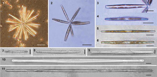
Figs 1–11. Light micrographs of Hyalosynedra lanceolata sp. nov. Figs 1–6. Colonies and chloroplasts in live cells. Figs 7–11. Cleaned specimens showing the lanceolate sternum and the wide cell size range. Scale: Figs 1–2 = 40 µm, Figs 3–6 = 20 µm, Figs 7–11 = 5 µm.
Figs 12–17. Scanning electron micrographs of Hyalosynedra lanceolata sp. nov. Fig. 12. Giant cells (200–300 µm long) with almost linear valves. Fig. 13. Internal view of valves, less than 100 µm long and with a lineal-lanceolate shape. Fig. 14. Colony of large cells and very small others showing the wide cell size range in culture. Figs 15–17. Cleaned specimens showing the lanceolate sternum. Scale = 5 µm, except Fig. 14 = 50 µm.
Figs 18–25. Scanning electron micrographs of Hyalosynedra lanceolata sp. nov. Fig. 18. Detail of the biseriate striation with alternate areolae and the lanceolate sternum at the centre of valves (up to one-third total width). Fig. 19. Apical part of cells showing areolae and a thin sternum. Fig. 20. Detail of the external openings of rimoportula and the rows of pores in ocellolimbus. Figs 21–22. Internal view of the smooth valves showing small rounded pores on both sides and the aperture of rimoportulae. Figs 23–24. Detail of valvocopula, copula and pleura with a row of pores (one apex in each image). Fig. 25. Cingular view of cells with the three girdle bands. Scale: Figs 18–24 = 1 µm, Fig. 25 = 10 µm.
Table 1. Diagnostic characters of Hyalosynedra lanceolata compared with other species of the genus.
Figs 26–34. Light and scanning electron micrographs of Synedra toxoneides from the Mar Menor lagoon. Fig. 26. Hyaline thin and curved cells under LM. Fig. 27. SEM micrographs of large curved cell with capitate poles. Figs 28–29. Detail of the visible striation under LM with immersion oil. Figs 30–31. External view of the wide sternum, striae and areolae. Fig. 32. Internal view of valves with rounded pores. Fig. 33. External view of rimoportula in a depressed area and a row of pores occupying the whole apex. Fig. 34. Internal opening of rimoportulae showing two parallel lips. Scale: Figs 26–27 = 20 µm, Figs 28–29 = 5 µm, Figs 30–34 = 1 µm.
3.3. Maximum likelihood tree inferred from a concatenated alignment of SSU rRNA and rbcL markers of 61 araphid diatoms. The numbers on the nodes are Bayesian posterior probability/maximum likelihood bootstraps (Bpp/MLb). The newly described species is shown in bold. Any support values lower than 50% were omitted.
Table 2. The AU test for the topologically unconstrained () and the four constrained trees in the monophyly analysis.