
Figures & data
Fig. 1. Map of Lake Ladoga and Lake Ilmen (north-western Russia) and the location of sampling points. Additional geographic information is provided in .
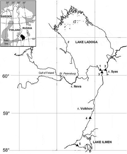
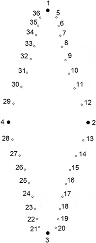
Fig. 2. Consensus configuration for Fragilaria heidenii and Staurosira tabellaria representing the placement of landmarks. Landmarks 1–4 are fixed. Semilandmarks 5–12 slide between 1 and 2. Semilandmarks 13–20 slide between 2 and 3. Semilandmarks 21–28 slide between 3 and 4. Semilandmarks 29–36 slide between 4 and 1.
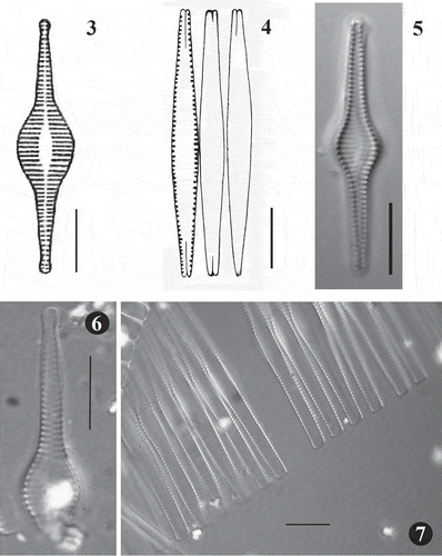
Figs 3–7. Fig. 3. Synedra inflata, original drawing produced by Heiden (1900). Fig. 4. Fragilaria heidenii, original drawing produced by Østrup (1910). Fig. 5. LM image of S. inflata from type slide made by Heiden (Rostock 0,15 Conventer See 3,2 m tief, B, Herbarium Berolinense). Fig. 6. LM image of broken valve of F. heidenii from original slide made by Østrup (3466, 20:190, C, Botanical Museum Copenhagen U096/2006 No. 1). Fig. 7. LM image of F. heidenii colonies in girdle view also from the slide made by Østrup. Scale = 10 µm.
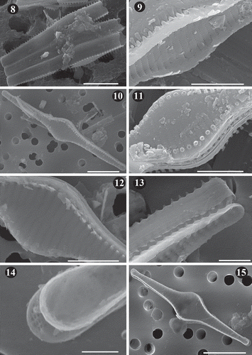
Figs 8–15. SEM images of Fragilaria heidenii Østrup from raw material collected by Østrup in Frederiksværk (Denmark) (C, Østrup Collection, Raw material 384.I Rant. 4244). Fig. 8. Broken frustules of a colony showing tight linkage between contiguous cells. Fig. 9. Inner view of central area showing details of the striae. Also, notice the spatulate spines linking neighbouring valves. Fig. 10. Outer view of valve showing striae, position of spines and lanceolate axial area. Fig. 11. Detail of central area in outer view showing the areolae on the mantle and broken off spines located on the interstriae. Fig. 12. Central area in outer view showing spatulate spines and small, elliptical to round areolae. Fig. 13. Side view of two neighbouring frustules showing unperforated girdle bands. Notice tapered spine structure toward valve apex and loose attachment between valves. Fig. 14. Inner view of apex covered by a film deposit. Notice structure of apical pore field on the valve below. Fig. 15. Inner view of valve. Scale = 10 µm (, , ), 5 µm (, , ), 2.5 µm (), 1 µm ().
Figs 16–19. SEM images of Fragilaria heidenii Østrup from raw material collected by Østrup in Frederiksværk (Denmark) (C, Østrup Collection, Raw material 384.I Rant. 4244). Fig. 16. Side view of two large contiguous valves. . Outer view of mid-region of valve showing lanceolate axial area, areolae varying from elliptical to round, location of spines and raised interstriae. Fig. 18. Inner view of mid-region of valve showing vola occlusion in areolae. Notice raised interstriae. Fig. 19. Middle part of two broken neighbouring frustules. Notice internal vola occlusion of areolae and striae extending onto the valve mantle. Scale = 4 µm (), 1 µm (–).
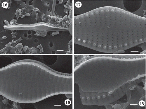
Table 1. General information of sites sampled for the present study.
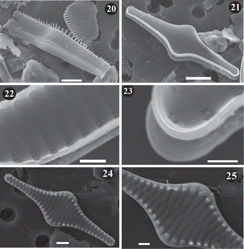
Figs 20–25. SEM images of Staurosira inflata. Lake Ladoga and Lake Ilmen material. Fig. 20. Side view of two neighbouring frustules showing unperforated girdle bands. Notice longer spines at the central part of the valve. Fig. 21. General internal view of frustule. Notice lack of rimoportulae. Fig. 22. Inner view of frustule. Notice vola occlusion of areolae and raised interstriae. Fig. 23. Inner view of apex. The small apical pore field can be seen. Fig. 24. General external view of frustule. Notice striae perpendicular to the apical axis at the valve centre and slightly radial at the ends. Fig. 25. Close up of valve in showing striae that extend onto the valve mantle. Scale = 5 µm (), µm (), 2 µm (), µm (, ), 0.5 µm ().
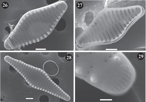
Figs 26–29. SEM images of Staurosira tabellaria. Lake Ladoga and Lake Ilmen material. Fig. 26. General external view of frustule showing details of striae and location of spines on the interstriae. Fig. 27. Internal view of frustule showing mantle at apexes. Notice the absence of rimoportulae and lack of ornamentation on girdle bands. Fig. 28. External view of large frustule. Notice raised interstriae and spines located on them. Fig. 29. Close up of apex of valve in . Notice details of apical pore field. Scale = 2 µm (–), 0.5 µm ().
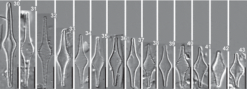
Figs 30–43. Staurosira inflata, Lake Ladoga and Lake Ilmen populations. LM photographs showing size diminution series. Scale = 10 µm.
Figs 44–58. Staurosira tabellaria, Lake Ladoga and Lake Ilmen populations. LM photographs showing size diminution series. Scale = 10 µm.
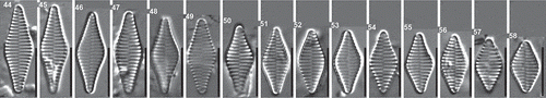
Table 2. Results of the analysis of covariance (ANCOVA), testing relationships among valve length and length-to-width ratio at different levels of categorical independent variables (species). Analysis tests whether the regression lines differ in their intercepts.
Fig. 59. Box-plot diagrams showing variation in valve length (A), width (B), length-to-width ratio (C), and striae density (D) of S. inflata (SI) and S. tabellaria (ST). Point represents mean, whiskers indicate max and min of the data.
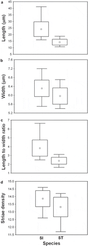
Fig. 60. Plot of length-to-width ratio against valve length for S. inflata and S. tabellaria. Each taxon is represented by linear regression line reflecting aspect ratio variation with length. The morphospace was contoured (dashed horizontal lines) by set of standard shape descriptors based on length-to-width ratios proposed by Cox (Citation1995). The ratios are shown on the left side of the plot; morphological characters (lanceolate, elliptical etc.) corresponding to these ratios are shown on the right side.
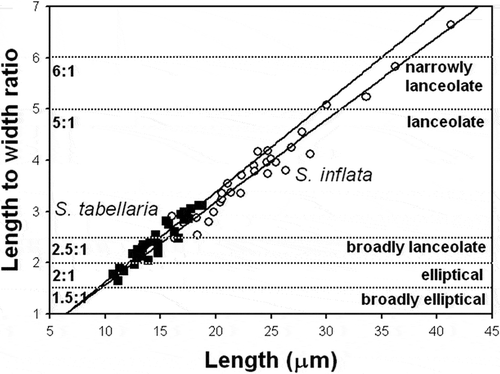
Table 3. Linear regression-derived parameters for relationships of relative warps to valve length of Staurosira heidenii and S. tabellaria.
Fig. 61. Plot of the first and second relative warps (RW1–2) for S. inflata and S. tabellaria. Group outliers are connected by lines. (a) S. inflata collected in Lake Ladoga and Lake Ilmen (north-western Russia); (b) S. inflata from raw material in the Østrup collection; (c) S. inflata from the Heiden’s type slide; (d) S. tabellaria collected in Lake Ladoga and Lake Ilmen; (e) S. tabellaria (syn: S. grigorszkyi) from raw material in the Pantocsek collection collected in Lake Balaton (Ács et al., 2009); (f) S. tabellaria (syn: S. grigorszkyi) from recent samples collected in Lake Balaton (Ács et al., 2009). Illustrations inside the plot represent shape variation of S. inflata and S. tabellaria in the morphospace of the first two relative warps (RW1–2).
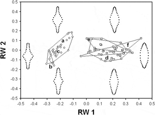
Fig. 62. Plot of the first relative warp (RW1) against valve length for S. inflata and S. tabellaria. Regression lines represent ontogenetic-allometric trends in S. inflata and S. tabellaria populations. Illustrations outside the plot represent extremes of the warp.
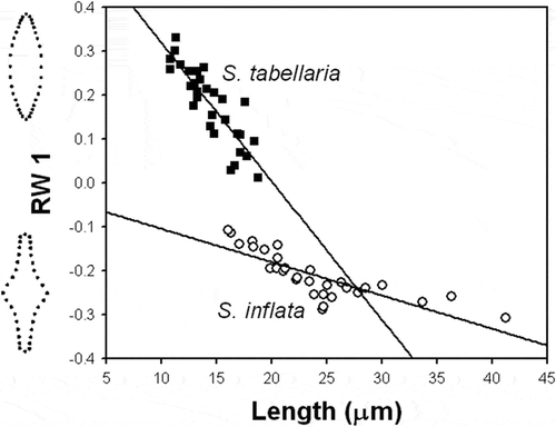
Fig. 63. Plot of the second relative warp (RW2) against valve length for S. inflata and S. tabellaria. Regression line represents ontogenetic-allometric trend in S. inflata population. Illustrations outside the plot represent extremes of the warp.
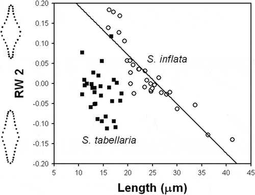
Table 4. Comparison of S. inflata with other morphologically similar taxa within the genus Staurosira.