Figures & data
Figs 1–2. Habit of Udotea geppiorum. Fig. 1. Typical habit of a mesophotic specimen collected from south-west O‘ahu Hawai‘i. Fig. 2. Specimen CS405A collected from 18 m depth in Guam. Note the conspicuous concentric segments on both specimens and the more relaxed/elongated holdfast and blade of the mesophotic specimen from Hawai‘i. Specimens display proportional size difference. Scale bars = 1 cm.
Figs 3–4. Blade anatomy of Udotea geppiorum. Fig. 3. Longitudinal section emphasizing the ‘tongue and groove’ arrangement of concentric segments and bridging medullary siphons between an inferior and a superior segment. Fig. 4. Longitudinal section emphasizing lateral branching of medullary siphons and the dense cortex. Scale bars = 50 μm.
Figs 5–9. Line drawings of Udotea geppiorum medullary siphons. Fig. 5. Blade siphon with lateral branchlets. Fig. 6. Distal blade siphon with lateral branchlets and chloroplasts. Fig. 7. Lateral branchlets from apical region of the blade. Fig. 8. Stipe lateral branchlet with chloroplasts. Fig. 9. Surface view of decalcified blade showing siphon apices. Scale bar = 30 μm.
Fig. 10. Bayesian majority-rule consensus tree depicting the phylogenetic position of Udotea geppiorum in the family Udoteaceae inferred by the chloroplast genes tufA. Values shown at nodes represent posterior probabilities scaled on 100% (before forward slash) and bootstrap support determined via Maximum likelihood analysis (after forward slash). Full support (> 99%) is noted by an asterisk (*) while values below 50% are indicated by an hyphen (-). Udotea spp. are displayed in bold with their sampling locality. Newly sequenced specimens of U. geppiorum are shaded in grey. Note the paraphyly of Udotea sensu stricto caused by the position of U. argentea, and the polyphyly of Udotea spp. sensu lato.
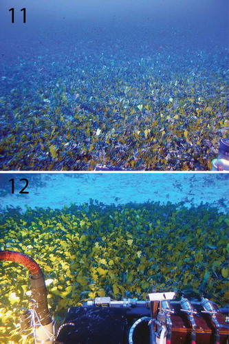
Figs 11–12. Submersible views of mesophotic meadows formed by Udotea geppiorum in south-west O‘ahu. Fig. 11. Overlooking an extensive meadow (Dive P4-188). Fig. 12. Edge of a meadow (Dive P5-606). Note that the meadows are so densely vegetated that the sand substrate is not visible among the blades. Picture taken by submersible pilot Terry Kerby in Kaiwi, O‘ahu.