
Figures & data
Table 1. Occurrence of Porphyra and Phycocalidia species along Indian coasts
Fig. 1. ML phylogenetic tree based on based on rbcL sequence data. Bootstrap values and aLRT (> 0.5) for ML analysis are shown at nodes
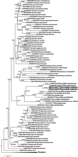
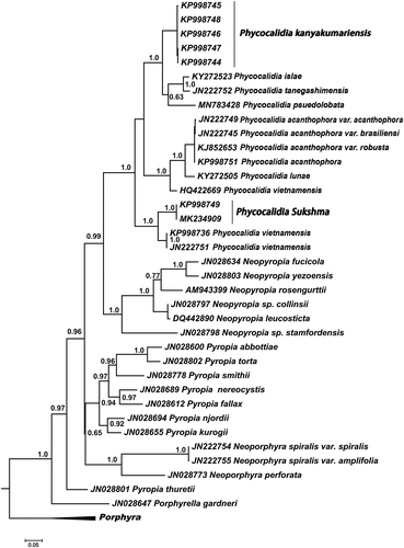
Fig. 2. ML phylogenetic tree based on COI-5P sequence data. aLRT (> 0.5) for ML analysis are shown at nodes
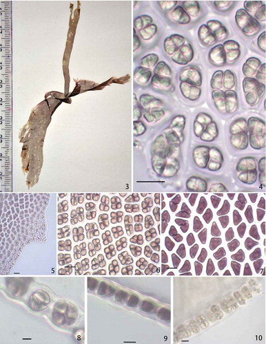
Figs 3–10. Habit and anatomy of Phycocalidia sukshma. Fig. 3. Habit. Fig. 4. Surface view of blade showing spermatangia. Fig. 5. Spinulose margin of the thallus. Fig. 6. Surface view of blade showing zygotosporangia. Fig. 7. Surface view of blade showing vegetative cells. Fig. 8. Cross-section of blade through zygotosporangia. Fig. 9. Cross-section of blade through vegetative cells. Fig. 10. Cross-section of blade through spermatangia. Scale bars = 10 µm
Figs 11–18. Habit and anatomy of Phycocalidia kanyakumariensis. Fig. 11. Habit of P. kanyakumariensis. Fig. 12. Spinulose margin of the thallus. Fig. 13. Cross-section of blade through zygotosporangia. Fig. 14. Cross-section of blade through spermatangia. Fig. 15. Surface view of blade showing vegetative cells. Fig. 16. Cross-section of blade through vegetative cells. Fig. 17. Surface view of blade showing zygotosporangia. Fig. 18. Surface view of blade showing spermatangia. Scale bars = 10 µm
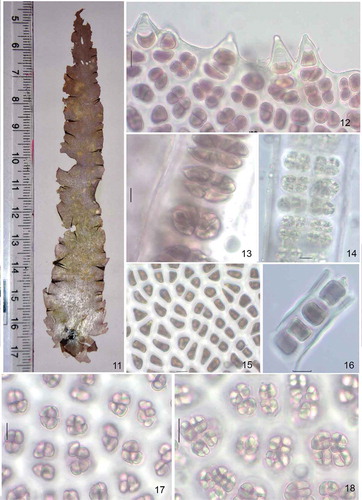
Table 2. Comparative morphological analysis of Phycocalidia species