547
Views
5
CrossRef citations to date
0
Altmetric
Original

Membrane interactions of peptides representing the polybasic regions of three Rho GTPases are sensitive to the distribution of arginine and lysine residues

, , , &
Pages 14-22 | Received 24 Jan 2007, Published online: 09 Jul 2009

Figures & data

Figure 1.  Aligned C-terminal sequences of the three human Rho protein family members Rac1, TCL and Cdc42, indicating (in upper case) the lysine- and arginine- rich polybasic region.

Figure 1.  Aligned C-terminal sequences of the three human Rho protein family members Rac1, TCL and Cdc42, indicating (in upper case) the lysine- and arginine- rich polybasic region.

Figure 2.  31P MAS NMR spectra of MLVs of DOPC/DOPG (2:1 molar ratio) at 4°C. Spectra are shown for the lipid sample alone (a) and after the addition of the 5 basic hexapeptides to a lipid/peptide molar ratio of 20:1 (b–f).
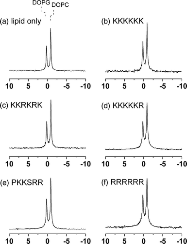
Figure 2.  31P MAS NMR spectra of MLVs of DOPC/DOPG (2:1 molar ratio) at 4°C. Spectra are shown for the lipid sample alone (a) and after the addition of the 5 basic hexapeptides to a lipid/peptide molar ratio of 20:1 (b–f).
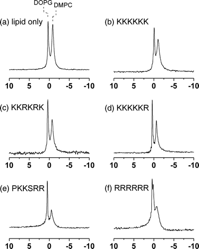
Figure 3.  31P MAS NMR spectra of MLVs of DMPC/DOPG (2:1 molar ratio) at 4°C. Spectra are shown for the lipid sample alone (a) and after the addition of the 5 basic hexapeptides to a lipid/peptide molar ratio of 20:1 (b–f).

Figure 3.  31P MAS NMR spectra of MLVs of DMPC/DOPG (2:1 molar ratio) at 4°C. Spectra are shown for the lipid sample alone (a) and after the addition of the 5 basic hexapeptides to a lipid/peptide molar ratio of 20:1 (b–f).

Table I.  Summary of 31P NMR isotropic chemical shifts (σi) and line widths at half height (Δν½) at 4°C for MLVs of DOPC/DOPG alone and in the presence of the 5 basic hexapeptides at a lipid/peptide molar ratio of 20:1.

Table II.  Summary of 31P NMR isotropic chemical shifts (σi) and line widths at half height (Δν½) at 4°C and apparent chemical shift anisotropy (Δσ) at 30°C for MLVs of DMPC/DOPG alone and in the presence of the 5 basic hexapeptides at a lipid/peptide molar ratio of 20:1.

Figure 4.  An illustration of the effect of lipid phase separation on the 31P MAS NMR spectra of DMPC/DOPG membranes. The diagram at the top illustrates the composition of the homogeneously mixed vesicles prepared from DMPC/DOPG in a 2:1 molar ratio (left) and from a mixture of preformed DMPC and preformed DOPG vesicles in the same ratio (right). Spectra corresponding to the two samples are shown underneath each diagram.
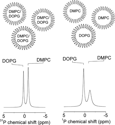
Figure 4.  An illustration of the effect of lipid phase separation on the 31P MAS NMR spectra of DMPC/DOPG membranes. The diagram at the top illustrates the composition of the homogeneously mixed vesicles prepared from DMPC/DOPG in a 2:1 molar ratio (left) and from a mixture of preformed DMPC and preformed DOPG vesicles in the same ratio (right). Spectra corresponding to the two samples are shown underneath each diagram.

Figure 5.  Wide-line 31P NMR spectra of MLVs of DMPC-d4/DOPG (2:1 molar ratio) at 30°C. Spectra are shown for the lipid sample alone (a) and after the addition of the 5 basic hexapeptides to a lipid/peptide molar ratio of 20:1 (bf).

Figure 5.  Wide-line 31P NMR spectra of MLVs of DMPC-d4/DOPG (2:1 molar ratio) at 30°C. Spectra are shown for the lipid sample alone (a) and after the addition of the 5 basic hexapeptides to a lipid/peptide molar ratio of 20:1 (b–f).
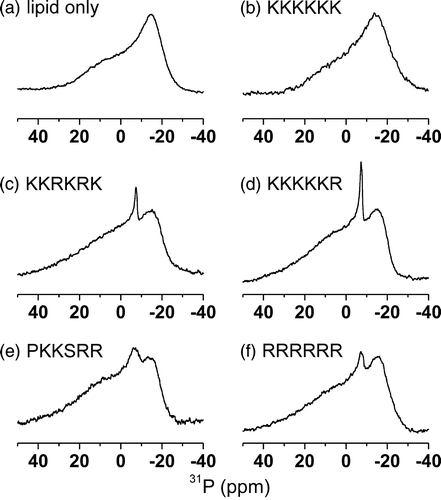
Figure 6.  Wide-line 2H NMR spectra of MLVs of DMPC-d4/DOPG (2:1 molar ratio) at 30°C. Spectra are shown for the lipid sample alone (a) and after the addition of the 5 basic hexapeptides to a lipid/peptide molar ratio of 20:1 (b–f). The quadrupole splittings for deuterons at the α- and β-choline positions of DMPC-d4 are denoted by the separation between the dotted lines.
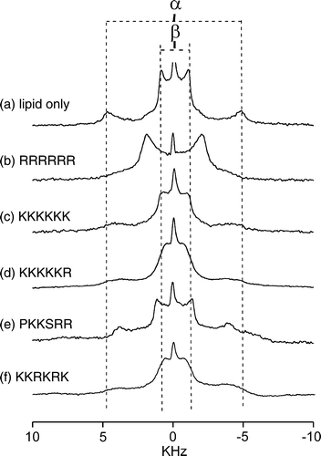
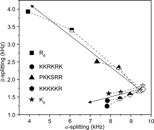
Figure 6.  Wide-line 2H NMR spectra of MLVs of DMPC-d4/DOPG (2:1 molar ratio) at 30°C. Spectra are shown for the lipid sample alone (a) and after the addition of the 5 basic hexapeptides to a lipid/peptide molar ratio of 20:1 (b–f). The quadrupole splittings for deuterons at the α- and β-choline positions of DMPC-d4 are denoted by the separation between the dotted lines.

Figure 7.  A plot of the 2H quadrupolar splittings (ΔνQ) at 30°C for the α- and β- deuterons of DMPC-d4 in the mixed DMPC-d4/DOPG membranes alone (open symbols) and after the addition of the basic hexapeptides to a lipid/peptide molar ratio of 50:1 (half-filled symbols) and 20:1 (full symbols).

Figure 7.  A plot of the 2H quadrupolar splittings (ΔνQ) at 30°C for the α- and β- deuterons of DMPC-d4 in the mixed DMPC-d4/DOPG membranes alone (open symbols) and after the addition of the basic hexapeptides to a lipid/peptide molar ratio of 50:1 (half-filled symbols) and 20:1 (full symbols).

Reprints and Corporate Permissions

Please note: Selecting permissions does not provide access to the full text of the article, please see our help page How do I view content?

To request a reprint or corporate permissions for this article, please click on the relevant link below:

Academic Permissions

Please note: Selecting permissions does not provide access to the full text of the article, please see our help page How do I view content?

Obtain permissions instantly via Rightslink by clicking on the button below:

If you are unable to obtain permissions via Rightslink, please complete and submit this Permissions form. For more information, please visit our Permissions help page.