Figures & data
Figure 1. The three-dimensional structure of the nAChR from Torpedo electric organ at 4Å resolution. This high resolution structure was obtained from electron images of helical tubes isolated from electric organ post-synaptic membranes from the marine ray Torpedo marmorata Citation[23]. These images were derived from the Protein Data Bank file 2BG9, coloured using the Swiss-Pdb Viewer, Deep View (www.expasy.org/spdbv) and rendered using MegaPov (www.povray.org). Individual subunits have been coloured for identification (α, red; β, blue; γ, green and δ, yellow). (A) A view from the extracellular side of the receptor. (B) A view from the side.
![Figure 1. The three-dimensional structure of the nAChR from Torpedo electric organ at 4Å resolution. This high resolution structure was obtained from electron images of helical tubes isolated from electric organ post-synaptic membranes from the marine ray Torpedo marmorata Citation[23]. These images were derived from the Protein Data Bank file 2BG9, coloured using the Swiss-Pdb Viewer, Deep View (www.expasy.org/spdbv) and rendered using MegaPov (www.povray.org). Individual subunits have been coloured for identification (α, red; β, blue; γ, green and δ, yellow). (A) A view from the extracellular side of the receptor. (B) A view from the side.](/cms/asset/3aba9190-524b-4997-bc16-398f75b64a96/imbc_a_303735_f0001_b.jpg)
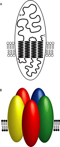
Figure 2. A model illustrating nAChR subunit topology and the pentameric arrangement of subunits in an assembled nAChR. A) A cartoon model of a nAChR subunit illustrating four transmembrane domains and the extracellular N- and C-termini. B) As discussed in the text, there is evidence that all assembled nAChRs (whether homomeric or heteromeric) are pentamers. One of the best characterized nAChRs is the heteromeric receptor expressed in the electric organ of the marine ray Torpedo. This representative cartoon image illustrates the known arrangement of subunits in the Torpedo electric organ nAChR. Individual subunits are illustrated by ovals and have been shaded to indicate the subunit type (α, red; β, blue; γ, green and δ, yellow).