Figures & data
Figure 1. Schematic diagram showing assembly and operation of a tangential flow filtration apparatus for bacterial membrane preparation. The lower panel shows a magnified view of the filter, to illustrate how membrane vesicles are separated from smaller, water-soluble proteins.
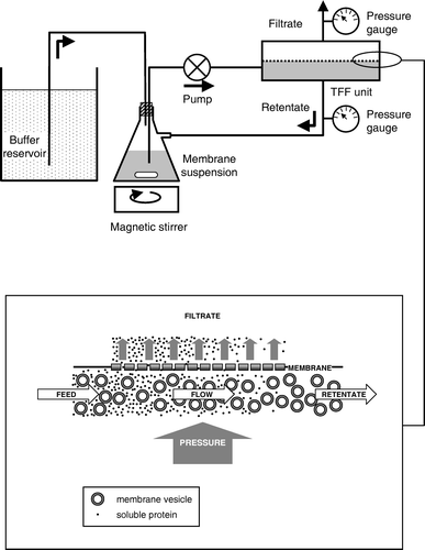
Figure 2. Flow diagram comparing the approximate timings of steps in bacterial membrane preparation by centrifugation and by tangential flow filtration. The points at which samples were taken for analysis are also indicated: S = supernatant, P = pellet, R = retentate, F = filtrate. During the 20 l wash stage of the tangential flow filtration procedure, filtrate samples were taken at 10 or 15 min intervals, and retentate samples at 15 or 30 min intervals, as detailed in .
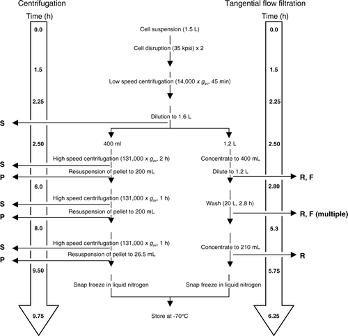
Figure 3. Time course of changes in the retentate and filtrate compositions during tangential flow filtration. (A) Protein concentration (•) in filtrate samples taken at the indicated times during the initial concentrating phase (0–15 min) and during the subsequent wash with 20 l buffer. (B) SDS-PAGE of filtrate samples (10 µl) taken at the indicated times, analysed for the presence of Npt1 by staining a western blot with antibody against the hexahistidine tag (upper panel) and for total protein by staining with SimplyBlue™ Safestain (lower panel). (C) Western blot of retentate samples (6.8 µg) taken at the indicated times, analysed for the presence of Npt1 by staining with antibody against the hexahistidine tag. The positions of marker proteins of known molecular mass, and of Npt1 (which migrates with an apparent molecular mass of 46 kDa), are shown on the left of B and C.
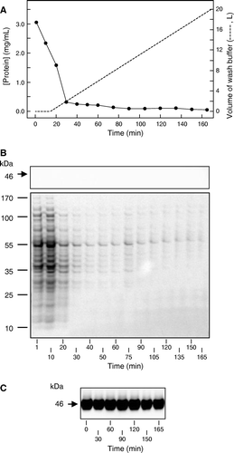
Table I. Comparison of centrifugal and tangential flow filtration membrane preparations.
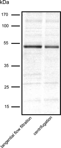
Figure 4. SDS-PAGE of Npt1 partially purified by Ni-NTA chromatography from DDM-solubilised membranes prepared either by centrifugation or tangential flow filtration, as indicated. Protein was detected by staining with SimplyBlue™ Safestain. The positions of marker proteins of known molecular mass are shown on the left.