2,661
Views
81
CrossRef citations to date
0
Altmetric
REVIEWS ON PROTEIN ACYLATION AND MICRODOMAINS IN MEMBRANE FUNCTION

Palmitoylation cycles and regulation of protein function (Review)

, PhD &
Pages 42-54 | Received 14 Nov 2008, Published online: 09 Jul 2009

Figures & data

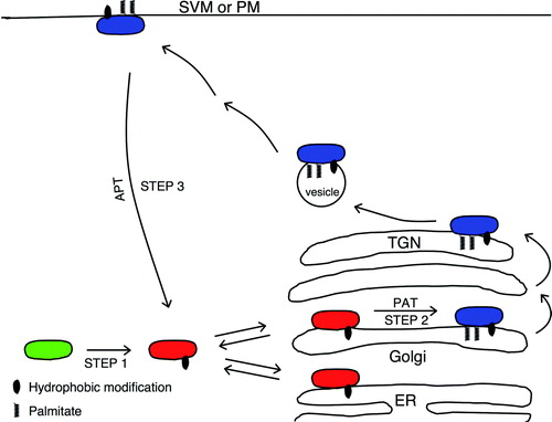
Figure 1.  Schematic model of cycling of the Ras and GAD65 proteins between cytosol, ER/Golgi, and PM/SVM. Newly synthesized hydrophilic and soluble protein (green) undergoes an irreversible hydrophobic modification in the cytosol (Step 1). The resulting hydrophobic protein (red) reversibly associates with ER and Golgi membranes, establishing an equilibrium between membrane and cytosolic pools. In Golgi membranes, the protein undergoes palmitoylation (Step 2) catalyzed by a palmitoyl-transferase. The palmitoylated protein (blue) is targeted to the TGN, shifting the equilibrium from ER and toward the Golgi. From the TGN, the protein is targeted to cytosolic vesicles and a pathway to either the PM (Ras) or synaptic vesicles (GAD65). A depalmitoylation along this pathway (Step 3) results in a reversal to the non-palmitoylated form, trafficking back to ER/Golgi membranes by a microtubule-independent pathway, and a new cycle of palmitoylation and targeting to a vesicular pathway. Bilayer membranes are shown as single lines for clarity. In reality, the lipid modifications interact only with the cytoplasmic leaflet of the bilayer. PAT, palmitoyl transferase; APT, acyl-protein thioesterase, ER, Endoplasmic reticulum; PM, plasma membrane; SVM synaptic vesicle membrane.

Figure 1.  Schematic model of cycling of the Ras and GAD65 proteins between cytosol, ER/Golgi, and PM/SVM. Newly synthesized hydrophilic and soluble protein (green) undergoes an irreversible hydrophobic modification in the cytosol (Step 1). The resulting hydrophobic protein (red) reversibly associates with ER and Golgi membranes, establishing an equilibrium between membrane and cytosolic pools. In Golgi membranes, the protein undergoes palmitoylation (Step 2) catalyzed by a palmitoyl-transferase. The palmitoylated protein (blue) is targeted to the TGN, shifting the equilibrium from ER and toward the Golgi. From the TGN, the protein is targeted to cytosolic vesicles and a pathway to either the PM (Ras) or synaptic vesicles (GAD65). A depalmitoylation along this pathway (Step 3) results in a reversal to the non-palmitoylated form, trafficking back to ER/Golgi membranes by a microtubule-independent pathway, and a new cycle of palmitoylation and targeting to a vesicular pathway. Bilayer membranes are shown as single lines for clarity. In reality, the lipid modifications interact only with the cytoplasmic leaflet of the bilayer. PAT, palmitoyl transferase; APT, acyl-protein thioesterase, ER, Endoplasmic reticulum; PM, plasma membrane; SVM synaptic vesicle membrane.

Table I.  Half-times of fluorescent protein recovery into the Golgi compartment following photobleaching.

Table II.  Intracellular localization of mammalian DHHC family of palmitoyl transferases.

Table III.  Mutations in DHHC proteins associated with pathology.
Reprints and Corporate Permissions

Please note: Selecting permissions does not provide access to the full text of the article, please see our help page How do I view content?

To request a reprint or corporate permissions for this article, please click on the relevant link below:

Academic Permissions

Please note: Selecting permissions does not provide access to the full text of the article, please see our help page How do I view content?

Obtain permissions instantly via Rightslink by clicking on the button below:

If you are unable to obtain permissions via Rightslink, please complete and submit this Permissions form. For more information, please visit our Permissions help page.