Figures & data
Figure 1. Structure of the isoprenoids farnesol (FN) and geranylgeraniol (GG).
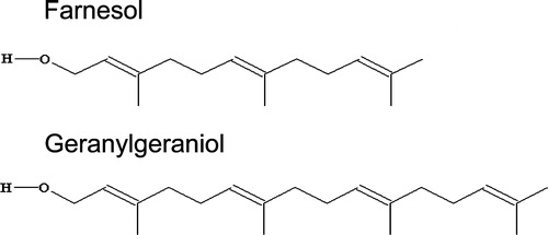
Figure 2. DSC heating (a, c) and cooling (b, d) thermograms of aqueous dispersions of 1,2-dielaidoyl-sn-glycero-3-phosphoethanolamine (DEPE) in absence and in presence of farnesol (FN) and geranygeraniol (GG) at the molar ratio indicated. The arrows (panel c) indicate the endothermic peak of the Lα-to-HII phase transition. The scan rate was 2°C/min. (e) Deconvolution of the calorimetric peak of DEPE:FN (20:1, mol:mol) corresponding to the Lβ-to-Lα transition (heating scan): experimental data (solid line), fitted overall (broken line) and individual components (dotted lines).
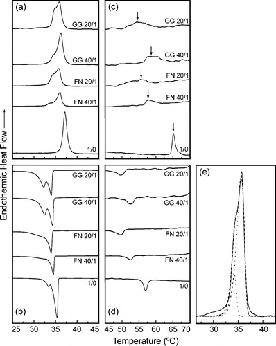
Table I. Calorimetric data of the effect of FN and GN in DEPE mixtures.
Figure 3. Sequence of SAXS (left) and WAXS (right) scattering patterns of (a, b) DEPE, (c, d) DEPE:FN (20:1, mol:mol) and (e, f) DEPE:GG (20:1, mol:mol). The scan rate was 1°C/min. Successive diffraction patterns were collected for 15 s every minute. The thermal sequence of the transition from (Lc + HII) to Lα and Lα-to-HII phases was clearly observed in the heating scan. A Lβ phase occur at the end of the cooling scan. The Lc and Lβ phases were identified by the reflections in the WAXS region. The Lα phase was identified by a single peak of reflection in the SAXS region with a very good signal-to-noise ratio. The HII phase was characterised by the appearance of three diffraction orders in the SAXS region with a d-spacing ratio of 1:1/√3:1/√4.
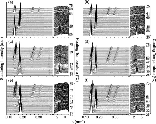
Figure 4. Sequence of scattering patterns from DEPE:FN (20:1, mol:mol). (a) In the heating scan, the transition from Lc to Lα is seen on the WAXS part of the scattering. Also the transition from Lc into HII (associated peaks marked by arrows) and Lα (sharp peaks) is evidenced. (b) In the cooling sequence, the transition from HII+Lc to Lα and the re-entrant HII co-existing with Lβ is shown.
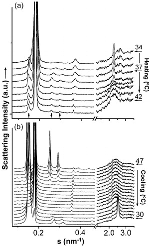
Figure 5. Dependence of the interplanar repeat distance (d) on temperature for (a) DEPE heating run, (b) DEPE:FN (40:1, mol:mol) heating run, (c) DEPE:FN (20:1, mol:mol) heating and (d) cooling run, (e) DEPE:GG (40:1, mol:mol) heating run and (f) DEPE:GG (20:1, mol:mol) heating run. The different phases represented are: Lc, Lβ, Lα and HII. The co-existence of the Lc + HII and Lα+HII phases is indicated by the interval temperature range covered by the vertical lines. The spacing corresponding to the first order of the re-entrant HII phase is not plotted for simplicity of the figure.
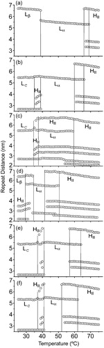
Table II. Summary of the structural properties.