Figures & data
Figure 1. Subcellular localization of ARFRP1-GFP and endogenous ARL1 in HeLa cells. HeLa cells were transfected with the cDNA of ARFRP1-Q79L-GFP, ARFRP1-T31N-GFP or ARFRP1-wt-GFP. Cells were fixed with methanol and stained for ARL1 with an affinity purified polyclonal antibody in combination with a TRITC-conjugated secondary antibody. Images of GFP-fluorescence and of TRITC were obtained by confocal laser scanning microscopy as described in Methods. This figure appears in colour in Molecular Membrane Biology online.
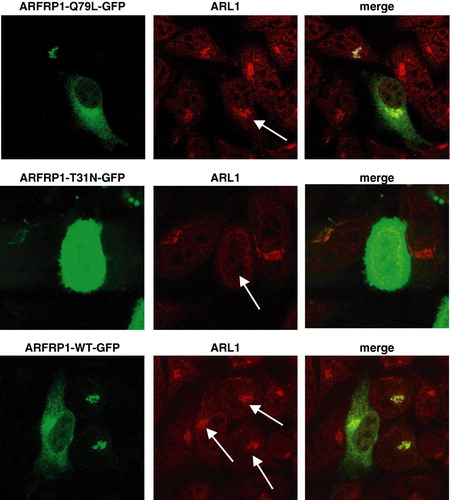
Figure 2. Subcellular localization of ARL1 (A), ARFRP1 (B), and Golgin-245 (A,B) in HeLa cells. HeLa cells were transfected with the cDNA of (A) ARL1-Q71L-GFP, ARL1-T31N-GFP, or (B) ARFRP1-Q79L-GFP or ARFRP1-T31N-GFP. Cells were fixed with methanol and stained for Golgin-245 with an affinity purified polyclonal antibody in combination with an Alexa546-conjugated secondary antibody. Images of GFP-fluorescence and of Alexa546 were obtained by confocal laser scanning microscopy as described in Methods. Arrows depict cells that expressed the respective ARL1 (A) or ARFRP1 (B) construct. This figure appears in colour in Molecular Membrane Biology online.
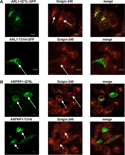
Figure 3. Silencing of the ARFRP1 gene in HeLa cells disrupts the Golgi association of GTP-bound ARL1. (A) Silencing of endogenous ARFRP1 in HeLa cells by small interference RNA. DNA encoding for small interference RNA (siRNA) targeting the human ARFRP1 gene was subcloned into the pSUPER vector and HeLa cells were transfected with the empty vector (−) or with the pSUPER-ARFRP1 construct (+), cells were lysed 4, 6, or 8 days after transfection and ARFRP1 expression was analysed by Western blotting. (B) Subcellular localization of ARL1 after silencing of ARFRP1 in HeLa cells. HeLa cells were transfected with pSUPER (control) or pSUPER-ARFRP1 (ARFRP1-siRNA). After 4 days, cells were transfected with cDNA of ARL1-Q71L-GFP or ARL1-T31N-GFP. Cells were fixed with methanol and the subcellular localization of the GFP-tagged ARL1 mutants was analyzed by confocal laser scanning microscopy as described in the Methods section. This figure appears in colour in Molecular Membrane Biology online.
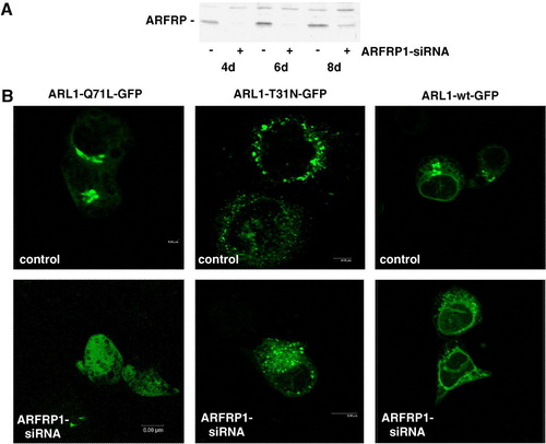
Figure 4. Inhibition of ARFRP1 expression in HeLa cells disrupts Golgi association of ARL1-Q71L (A), and endogenous ARL1 (B) and subsequently of Golgin-245 (A, B). (A) ARFRP1 expression was downregulated in HeLa cells by transfection of pSUPER-ARFRP1. After 4 days, cells were transfected with cDNA of ARL1-Q71L-GFP. Cells were fixed with methanol and stained for Golgin-245 with a polyclonal antibody in combination with an Alexa546-conjugated secondary antibody. (B) ARFRP1 expression was downregulated in HeLa cells by transfection of pSUPER-ARFRP1. Control cells were transfected with scrambled siRNA. Four days after transfection, cells were fixed with methanol, and endogenous ARL1 was stained with an affinity-purified polyclonal anti-ARL1 antibody in combination with an Alexa488-conjugated secondary antibody. Golgin-245 was visualized with a polyclonal anti-Golgin-245 antibody in combination with an Alexa546-conjugated secondary antibody. Subcellular localization of the GFP-tagged ARL1-Q71L, of endogenous ARL1, and Golgin-245 was analysed by confocal laser scanning microscopy as described in the Methods. Arrows depict cells with disrupted Golgi association of ARL1 and Golgin-245.
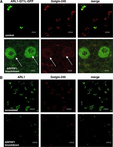
Figure 5A.
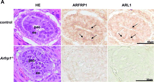
Figure 5. ARL1 targeting is disrupted in Arfrp1−/ − embryos. Immunohistochemical detection of ARFRP1 and ARL1 in control and Arfrp1−/ − embryos at day E 6.5 (A) and of fat tissue associated with the uterus of Arfrp1+/ − mother (B). Serial transversal sections of uteri at the stage of E6.5 were prepared and stained with hematoxilin and eosin (HE) or used for immunohistochemical detection of ARFRP1 and ARL1. ee, embryonic ectoderm; pac, proaminotic cavity.
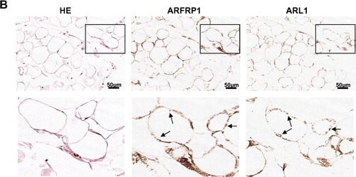
Figure 6. Inhibition of ARFRP1 expression in HeLa cells does not alter the structure of the cis-Golgi. ARFRP1 expression was downregulated in HeLa cells by transfection of pSUPER-ARFRP1 and 4 days later cells were fixed with methanol and (A) stained for ARL1 with an affinity purified polyclonal anti-ARL1 antibody in combination with an Alexa488-conjugated secondary antibody and co-stained for GM130 with a monoclonal antibody in combination with an Alexa546-conjugated secondary antibody or (B) stained for Golgin-245 with a polyclonal antibody in combination with an Alexa488-conjugated secondary antibody and co-stained for giantin with a monoclonal antibody in combination with an Alexa546-conjugated secondary antibody. Immunofluorescence was analysed by confocal laser scanning microscopy as described in the Methods.
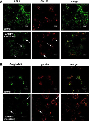
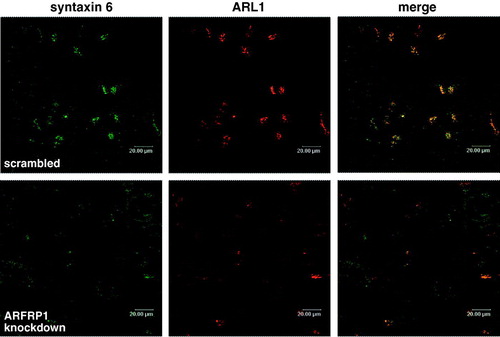
Figure 7. Depletion of ARFRP1 expression in HeLa cells alters TGN structure and dislocates syntaxin 6. HeLa cells were transfected with scrambled siRNA or with ARFRP1-specific siRNA and 4 days later cells were fixed with 4% paraformaldehyde and stained for ARL1 in combination with an Alexa546-conjugated secondary antibody and for syntaxin 6 in combination with an Alexa488-conjugated secondary antibody. Immunofluorescence was analysed by confocal laser scanning microscopy as described in the Methods.