Figures & data
Figure 1. Gravid uterus of rabbit; both branches of uterine artery have been closed for 3 and 9 minutes; colour changed but no remarkable change was observed from the third minute.
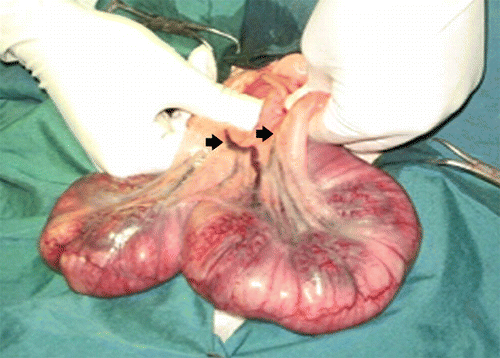
Table 1. Number of rabbit's neonates with different intensity of hypoxia in study groups based on clinical findings.
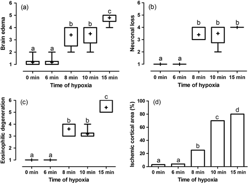
Figure 2. Box and whisker plot of median, mean and range of changes in brain oedema (a), neuronal loss (b), eosinophilic degeneration (c) and mean of percent of ischemic cortical area (d) of brain sampled of rabbit's neonates with different intensity of hypoxia. Different superscript letters indicates significant statistical difference (p<0.05) between groups using Mann–Whitney U test. Score 1 means 0–4%; score 2, 5–24%; score 3, 25–49%; score 4, 50–74%; score 5, 75–99%; and score 6, 100% histopathologic changes.