Figures & data
Figure 1. Skull, undifferentiated sarcoma of a dog showing proliferation of anaplastic and pleomorphic cells with a fusiform to epithelioid shape with moderate eosinophilic cytoplasm. H&E×40.
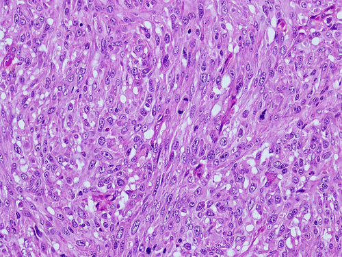
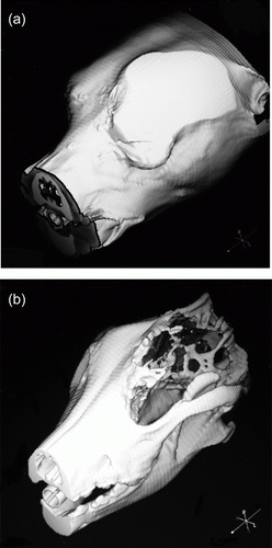
Figure 2. Three-dimensional reconstructed CT image of the skull. (a) Lateral reconstruction of the head where a mass formation occupying the orbital fossa is visualised. (b) 3D image with extraorbital involvement and focused on frontal and parietal areas, showing bone lysis and invasion into the cranial cavity.