2,787
Views
3
CrossRef citations to date
0
Altmetric
Original Articles

Histomorphological and histochemical studies on the different layers of skin of Bakerwali goat

, , &
Pages 208-213 | Received 27 Sep 2013, Accepted 27 May 2014, Published online: 08 Oct 2014

Figures & data

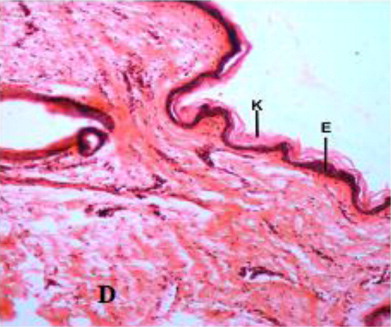
Figure 1. Skin of Bakerwali goat showing; keratin (K), epidermis (E) and dermis(D) in H&E X 100 in neonatal neck dorsal region.

Table 1. Total thickness of skin (µm) at different post-natal age groups and in different regions of Bakerwali goat (Mean ± SE).
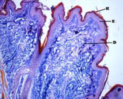
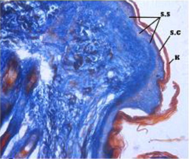
Figure 2. Skin of Bakerwali goat showing; keratin (K), epidermis (E) and dermis(D) in H&E X 100 and in Ayoub Schklar stain X in neonatal neck dorsal region.
Figure 3. Stratum spinosum (S.S), stratum cornium (S.C), stratum basali, epidermis and dermis and Keratin layer in Ayoub Schklar X 200.
Figure 4. Stratum spinosum (S.S), stratum cornium (S.C), stratum basali, epidermis and dermis and Keratin layer in Ayoub Schklar X 200.
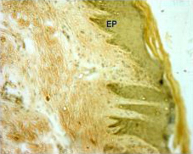
Figure 5. Skin of Bakerwali goat showing; epidermal pegs (EP) in Von Gieson and Verhoeff's X 100.
Figure 6. Thick epidermis (E) and typical epidermal pegs (EP) in thorax ventral region in H&E X 100.

Table 2. Thickness of epidermis (µm) at different post-natal age groups and in different regions of Bakerwali goat (Mean ± SE).
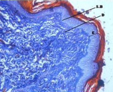
Figure 7. Epidermis(E), Dermis(D) and Presence of hair follicle (HF), Arrector pilli muscles (AP) in dermis in Masson's Tricrome stain X 100.
Figure 8. Keratin (K), Epidermis (E), papillary dermis (PD) and Reticular dermis (RD) layers in loin dorsal region in H&E X 100.

Table 3. Thickness of dermis (µm) at different post-natal age groups and in different regions of Bakerwali goat (Mean ± SE).
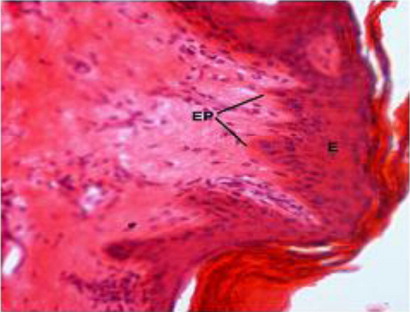
Table 4. Thickness of papillary dermis (µm) at different post-natal age groups and in different regions of Bakerwali goat (Mean ± SE).
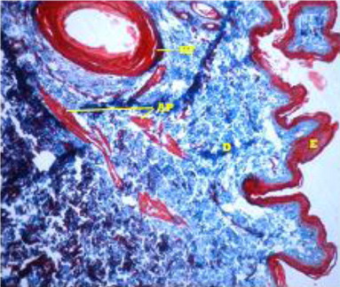
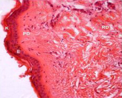
Table 5. Thickness of reticular dermis (µm) at different post-natal age groups and in different regions of Bakerwali goat (Mean ± SE).

Reprints and Corporate Permissions

Please note: Selecting permissions does not provide access to the full text of the article, please see our help page How do I view content?

To request a reprint or corporate permissions for this article, please click on the relevant link below:

Academic Permissions

Please note: Selecting permissions does not provide access to the full text of the article, please see our help page How do I view content?

Obtain permissions instantly via Rightslink by clicking on the button below:

If you are unable to obtain permissions via Rightslink, please complete and submit this Permissions form. For more information, please visit our Permissions help page.