Figures & data
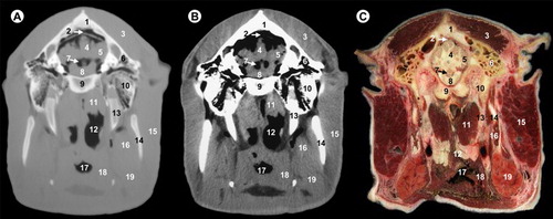
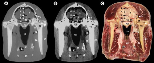
All views are rostral. 1, frontal sinus; 2, cribriform plate of ethmoid bone; 3, frontal cortex; 4, olfactory bulb; 5, sphenoidal sinus; 6, extraperiorbital fat; 7, zygomatic process of temporal bone; 8, body of presphenoid bone; 9, vomer bone; 10, tensor and levator palatine muscles; 11, pars nasalis pharyngis; 12, perpendicular plate of palatine bone; 13, upper third molar; 14, soft palate; 15, masseter muscle; 16, oral cavity; 17, lower third molar; 18, tongue and lingual fat; 19, hyoid bone; 20, mandibular canal.
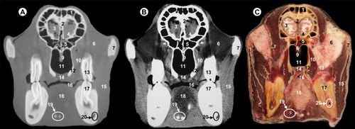
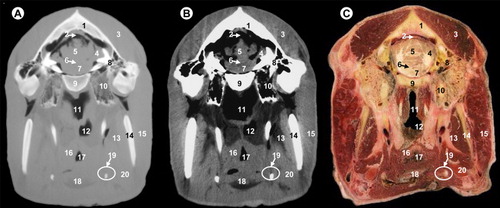
All views are rostral. 1, parietal bone; 2, dorsal sagittal sinus; 3, cerebral longitudinal fissure; 4, temporal muscle; 5, squamous part of temporal bone; 6, cerebral hemisphere; 7, coronoid process of mandible; 8, wing of presphenoid bone; 9, body of presphenoid bone; 10, cavernous sinus; 11, zygomatic process of temporal bone; 12, vomer bone; 13, pars nasalis pharyngis; 14, lateral pterygoideus muscle; 15, perpendicular plate of palatine bone; 16, ramus of mandible; 17, masseter muscle; 18, soft palate; 19, pharyngeal muscles; 20, pars oralis pharyngis; 21, tongue and lingual fat; 22, hyoid bone; 23, mandibular canal; 24, medial pterygoideus muscle.
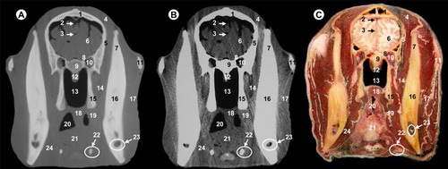
All views are rostral. 1, parietal bone; 2, dorsal sagittal sinus; 3, cerebral longitudinal fissure; 4, lateral ventricle; 5, cerebral hemisphere; 6, temporal muscle; 7, squamous part of temporal bone; 8, third ventricle; 9, thalamus; 10, hypothalamus; 11, hypophysis; 12, cavernous sinus; 13, body of basisphenoid bone; 14, zygomatic process of temporal bone; 15, temporomandibular joint; 16, maxillary vein; 17, lateral pterygoideus muscle; 18, pars nasalis pharyngis; 19, ramus of mandible; 20, mandibular canal; 21, masseter muscle; 22, medial pterygoideus muscle; 23, pharyngeal muscles; 24, pars oralis pharyngis; 25, hyoid bone.
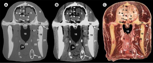
All views are rostral. 1, parietal bone; 2, dorsal sagittal sinus; 3, temporal muscle; 4, cerebral longitudinal fissure; 5, cerebral hemisphere; 6, third ventricle; 7, mesencephalic tectum; 8, mesencephalic aqueduct; 9, temporal sinus; 10, cerebral peduncle; 11, subarachnoid space; 12, body of basisphenoid bone; 13, ophthalmic, abducens, trochlear and oculomotor nerves; 14, zygomatic process of temporal bone; 15, temporomandibular joint; 16, condylar process of mandible; 17, lateral pterygoideus muscle; 18, maxillary vein; 19, pars nasalis pharyngis; 20, medial pterygoideus muscle; 21, masseter muscle; 22, hyoid bone; 23, pharyngeal muscles; 24, pars oralis pharyngis; 25, epiglottis.
All views are rostral. 1, parietal bone; 2, transverse sinus; 3, temporal muscle; 4, cerebellar hemisphere; 5, cerebellar vermis; 6, fourth ventricle; 7, pons; 8, petrous part of temporal bone; 9, basilar part of occipital bone; 10, tympanic part of temporal bone; 11, pars nasalis pharyngis; 12, pars laryngea pharyngis; 13, medial pterygoideus muscle; 14, ramus of mandible; 15, parotid gland and masseter muscle; 16, pharyngeal muscles; 17, vestibule of larynx; 18, epiglottis; 19, hyoid bone; 20, mandibular gland.
All views are rostral. 1, parietal bone; 2, transverse sinus; 3, temporal muscle; 4, cerebellar vermis; 5, cerebellar hemisphere; 6, temporal sinus; 7, fourth ventricle; 8, pons; 9, basilar part of occipital bone; 10, petrous part of temporal bone; 11, rectus ventralis and longus capitis muscles; 12, pars laryngea pharyngis; 13, hyoid bone; 14, ramus of mandible; 15, parotid gland and masseter muscle; 16, medial pterygoideus muscle; 17, vestibule of larynx; 18, pharyngeal muscles; 19, mandibular gland.