Figures & data
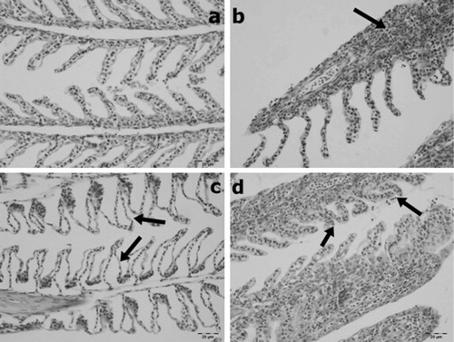
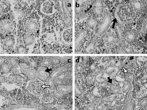
Table 1. Time- and dose-dependant comparison of histopathology in gill tissue.
Table 2. Time- and dose-dependant comparison of histopathology in kidney tissue.
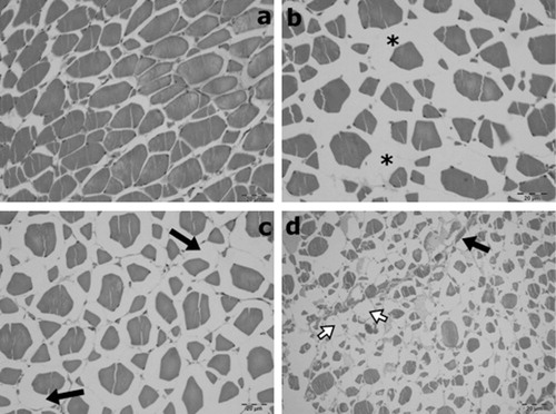
Table 3. Time- and dose-dependant comparison of histopathology in muscle tissue.