Figures & data
Figure 1. Ultrasonographic appearance of affected left ovary. On D5, the left ovary revealed mass with solid appearance (a). On D22, the left ovary become enlarged and merged multicystic appearance (b). Bar = 10 mm.
Figure 2. Gross appearance of the extracted left ovary. The extracted left ovary (60 mm × 70 mm × 70 mm, 117 g in weight) was coated smooth surface (a). The cut surface revealed many various-sized cystic follicles filled with yellowish serous fluids and greyish-yellow solid parts (↑) (b).
Figure 3. Microscopic appearance of the section of the extracted left ovary. The tissues were observed as various-sized follicular structures contained eosinophilic-fluid-like Call-Exner body (a: ×100). The neoplastic cells in parts of the tumour were also formed with tubular and solid patterns (b: ×200).
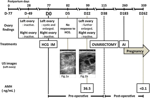
Figure 4. Clinical course of the present case after parturition. Note: D0: day of first medical examination; US images: ultrasound images.