932
Views
2
CrossRef citations to date
0
Altmetric
Articles

Electrocardiographic dynamic development and heart rate variability in lambs during the neonatal period

, ORCID Icon, , ORCID Icon, , ORCID Icon, & ORCID Icon show all
Pages 1137-1143 | Received 25 Oct 2017, Accepted 20 Feb 2018, Published online: 15 May 2018

Figures & data

Figure 1. Placement of ‘alligator’-type electrodes and manual restraint in right latero-lateral decubitus for performing electrocardiography in the frontal plane.
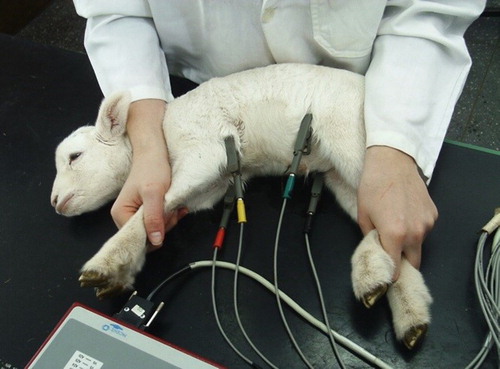
Figure 1. Placement of ‘alligator’-type electrodes and manual restraint in right latero-lateral decubitus for performing electrocardiography in the frontal plane.

Figure 2. Electrocardiographic tracing of lamb 20, deriving DII, frontal, and birth (24 h) at 35 days of age (25 mmseg, N).
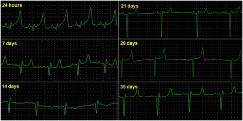
Figure 2. Electrocardiographic tracing of lamb 20, deriving DII, frontal, and birth (24 h) at 35 days of age (25 mmseg, N).

Table 1. Mean and standard deviation of variables obtained by ECG in the frontal plane at the second moment of evaluation.

Table 2. Mean and standard error* of variables obtained by 24-h ECG using a Holter system for the second moment of evaluation.