Figures & data
Figure 1. Skin lesion development of TST in the guinea pigs infected with Mtb by drinking water. A: Guinea pig receiving high dose of Mtb (2 × 10⁷ CFU) group; B: Guinea pig receiving low dose of Mtb (2 × 10⁵ CFU) group. Arrow a as a negative control and Arrow b representative images of lesions at 4 weeks post-infection.
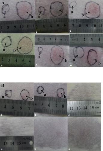
Figure 2. Body weight of guinea pigs measured during the study period. After challenged with 2 × 10⁷ CFU of Mtb, guinea pigs gained less weight compared to the uninfected control. Change in body weight is displayed as the mean of weights of two groups per time in this graph.
Table 1. Effects of Mtb infected on body weight gain in the guinea pigs.
Figure 3. Histopathologic changes in selected tissues from infected guinea pigs. (A,B) Lung section of guinea pigs infection with 2 × 10⁷ CFU of Mtb. Necrotic granulomas (arrow) were observed in the tissues from both right and left lung lobes at 20 weeks post-infection (×4). (C) Caseous necrosis in the centre of the lesions developed at 20 weeks post-infection, and the necrotic granuloma was confined by surrounding fibroblast (arrow) and fibrocyte in the spleen (×4). (D) Severe necrotizing fibrous granulomas were also appeared in the mesenteric lymph glands, also the necrotic granuloma was confined by surrounding fibroblast (arrow) and fibrocyte (×4).
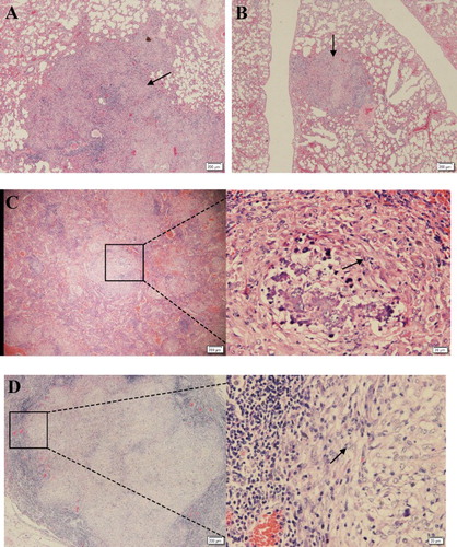
Figure 4. Bacterial burden in guinea pigs infected via route of drinking water containing 2 × 10⁷ CFU of Mtb.