Figures & data
Table 1. Localization and prevalence of lesions specific for contact dermatitis, P < 0.01.
Figure 1. Moderate to strong swelling affecting the metatarsal region due to perifocal inflammatory oedema in plantar pododermatitis.
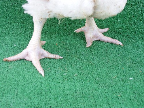
Figure 2. Plantar pododermatitis lesions, having undergone maceration. Marked dehydration emerging on tarsometatarsal aspects of legs (arrows) likely to prolonged lying down and inability to reach food and water.
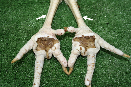
Figure 3. Plantar pododermatitis lesion exposed after removal of faecal mass and litter stuck on it. Well-demarcated crater-like ulcers surrounded by sharply demarcated haemorrhagic zone.
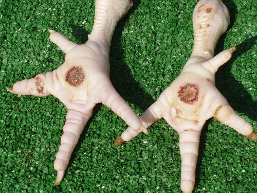
Figure 4. Breast, skin lesion of an erosive necrotic type in a 38-day broiler chicken after processing at the slaughterhouse.
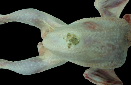
Figure 5. Contact dermatitis in the region of tarsometatarsal joints. Affected skin is necrotic, and of dirty grey colour.
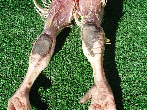
Table 2. aExtent and area of observed contact dermatitis lesions, P < 0.01.
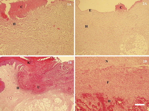
Figure 6. Histological structure of lesions in different stages of contact dermatitis. (1) Plantar lesions: 1A – a superficial lesion manifesting itself in an erosive skin defect, covered by crustose detritus (C) and heterophilic infiltration (H) in the underlying layers; 1B – a deep ulcerative lesion exhibiting defective keratinization in Str. intermedium (K), especially around the ulcer (U) and heterophilic infiltration (H) of the adjacent epidermis. (2) Breast lesions: 2A – an erosive lesion (E) affecting the epidermal layer, remainders of crustose detritus on the surface and heterophilic infiltration (H) in the underlying layers (C); 2B – advanced stage of organizing a lesion defect manifesting itself in the growth of fibrous tissue (F), from the destructive surface (D), which replaces the deposited necrotic detritus (N), H/E, Bar = 30 µm.