Figures & data
Table 1. Mean of body weights of control and VNDV-infected birds (g) ± SEM.
Table 2. Comparison of the cumulative mortality rate of broiler and pullets challenged with vvNDV.
Figure 1. (A) Proventriculus of VNDV infected broilers showing severe mucosal haemorrhages on day 4 PI. (B) Proventriculus of VNDV infected pullets showing mild mucosal haemorrhages on day 4 PI.
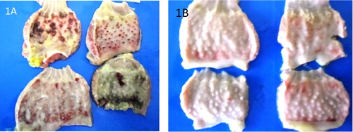
Figure 2. (A) Intestines of VNDV infected broilers showing severe haemorrhagic ulcers in broilers on day 4 PI. (B) Intestines of VNDV infected pullets showing less severe ulcers on day 4 PI.
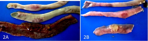
Figure 3. (A) Caecum of VNDV infected broiler showing severe swollen hemorrhagic tonsil on day 4 PI. (B) Caecum of VNDV infected pullet showing less severe lesion on the tonsil on day 4 PI.
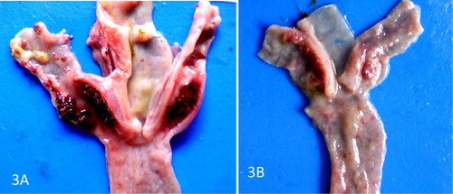
Figure 4. (A) Severe atrophy of the bursa of VNDV infected broilers on day 5 PI. (B) Less severe atrophy of the bursa of VNDV infected pullets on day 5 PI.
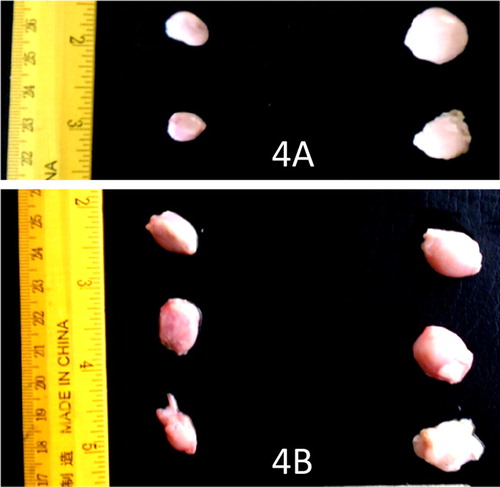
Figure 5. (A) Severe necrosis and depletion of lymphocytes in the bursa of VNDV infected broilers on day 4 PI. H&E × 400. (B) Less severe necrosis and depletion of the lymphocytes in the bursa of VNDV infected pullets on day 4 PI H&E × 400.
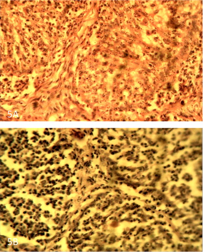
Figure 6. (A) Thymus of VNDV infected broiler showing severe necrosis and depletion of lymphocytes on day 4 PI. H&E × 400. (B) Thymus of VNDV infected pullet showing less severe necrosis and depletion of lymphocytes on day 4 PI. H&E × 400.
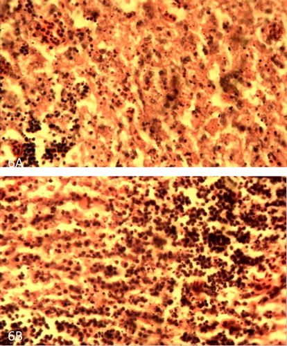
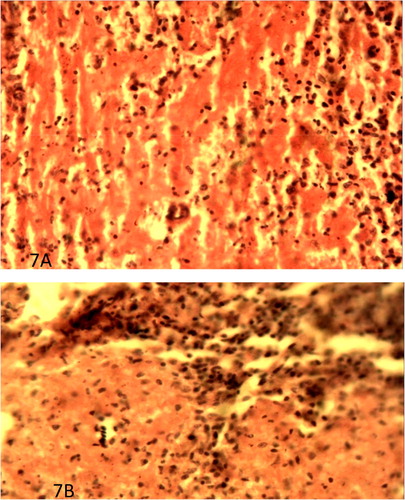
Figure 7. (A) Spleen of VNDV infected broiler showing severe lymphocytic depletion and fibrin deposition on day 4 PI. H&E × 400. (B) Spleen of VNDV infected pullet showing less severe lymphocytic depletion and fibrin deposition on day 4 PI. H&E × 400.