Figures & data
Table 1. Primers used for qRT-PCR.
Figure 1. Relative expression values of Th1 cytokines (IL-2, IFN-γ and TNF-β) and Th2 cytokines (IL-4, IL-5, IL-6 and IL-10) mRNA in spleens of nonpregnant and pregnant ewes measured by quantitative real-time PCR.
Note: DN16 = Day 16 of the oestrous cycle; DP13 = Day 13 of pregnancy; DP16 = Day 16 of pregnancy; DP25 = Day 25 of pregnancy. Significant differences (P < 0.05) are indicated by different letters within same colour column.
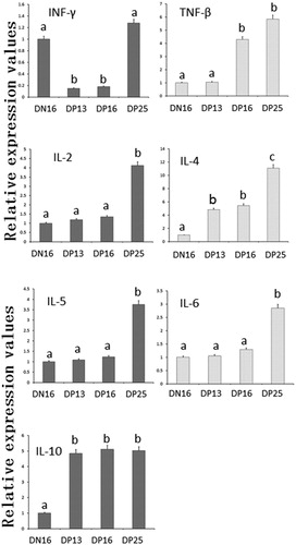
Figure 2. Expression of Th1 cytokines (IL-2, IFN-γ and TNF-β) and Th2 cytokines (IL-4, IL-5, IL-6 and IL-10) proteins in spleens of nonpregnant and pregnant ewes analyzed by western blot.
Note: hTNF-β = Recombinant human TNF-β protein; hIL-5 = Recombinant human IL-5 protein; hIL-10 = Recombinant human IL-10 protein; DN16 = Day 16 of the oestrous cycle; DP13 = Day 13 of pregnancy; DP16 = Day 16 of pregnancy; DP25 = Day 25 of pregnancy. Significant differences (P < 0.05) are indicated by different superscript letters within the same colour column.
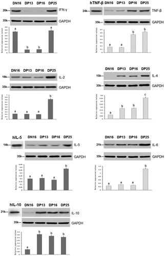
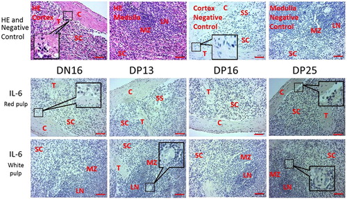
Figure 3. Representative immunohistochemical localization of IL-6 protein in spleens of nonpregnant and pregnant ewes. The spleen is divided into red pulp and white pulp, and surrounded by a thickened capsule. The red pulp and white pulp are not separated completely in this figure. Capsule (C) with several trabeculae (T) projects into the substance of the spleen.
Note: HE = stained by hematoxylin and eosin; SS = splenic sinuses; SC = splenic cords; MZ = marginal zone; LN = lymphoid nodule; DN16 = day 16 of the oestrous cycle; DP13 = day 13 of pregnancy; DP16 = day 16 of pregnancy; DP25 = day 25 of pregnancy. Bar = 50 µm.