Figures & data
Table 1. Primer sequences of MyHCs and 18S rRNA used in qPCR.
Figure 1. Histological structure of muscle fibres in Mashen and Large White pigs.
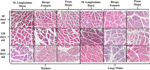
Figure 2. Muscle fibre diameter of the m. longissimus thoracis (a), biceps femoris (b), and psoas major (c) in Mashen and Large White pigs at different developmental stages. Comparison of muscle fibre diameters of the three skeletal muscles in Mashen pigs (d) and Large White pigs (e). (a–c): In different stages of Mashen pig, different lowercase letters indicate significant difference, while the same lowercase letters indicate no significant difference; In different stages of Large White pig, different capital letters indicate significant difference, while the same capital letters indicate no significant difference. * indicates that there are significant differences between Mashen and Large White pigs at the same stage. (d,e): In different tissues of the same age, different lowercase letters mean significant difference, while the same lowercase letters mean no significant difference.
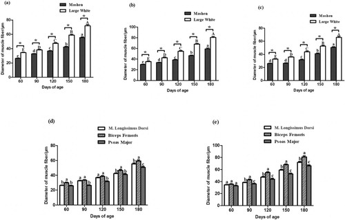
Figure 3. The developmental expression patterns of MyHCs in Mashen and Large White pigs at different developmental stages. The developmental expression patterns of MyHCI (a), MyHCIIa (d), MyHCIIx (g) and MyHCIIb (j) in m. longissimus thoracis. The developmental expression patterns of MyHCI (b), MyHCIIa (e), MyHCIIx (h) and MyHCIIb (k) in biceps femoris. The developmental expression patterns of MyHCI (c), MyHCIIa (f), MyHCIIx (i) and MyHCIIb (l) in psoas major. Note: In different stages of Mashen pig, different lowercase letters indicate significant difference, while the same lowercase letters indicate no significant difference; In different stages of Large White pig, different capital letters indicate significant difference, while the same capital letters indicate no significant difference. * indicates that there are significant differences between Mashen and Large White pigs at the same stage.
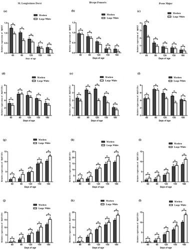
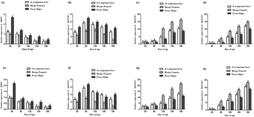
Figure 4. The expression patterns of MyHCs in different skeletal muscles. The expression patterns of MyHCI (a), MyHCIIa (b), MyHCIIx (c) and MyHCIIb (d) in Mashen pigs; (e-h) The expression patterns of MyHCI (e), MyHCIIa (f), MyHCIIx (g) and MyHCIIb (h) in Large White pigs. Note: In different tissues of the same age, different lowercase letters mean significant difference, while the same lowercase letters mean no significant difference.