Figures & data
Figure 1. Anatomical position of the viscera in a camel at postmortem examination. Image (a) shows a right-sided view of the liver, right lung, cranioventral ruminal sac, omasum, abomasum and intestines. Image (b) shows a left-sided view for the left lung, rumen, spleen and intestines. 1 = glandular sac of the caudodorsal ruminal sac; 2 = cranioventral ruminal sac.
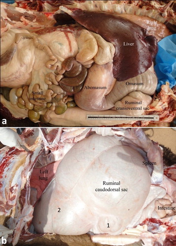
Figure 2. Anatomy of the interior of the rumen with smooth mucus membrane (a). Image (b) shows a close-up view for the ruminal glandular sacs. 1 = smooth ruminal mucosa; 2 = glandular sacs.
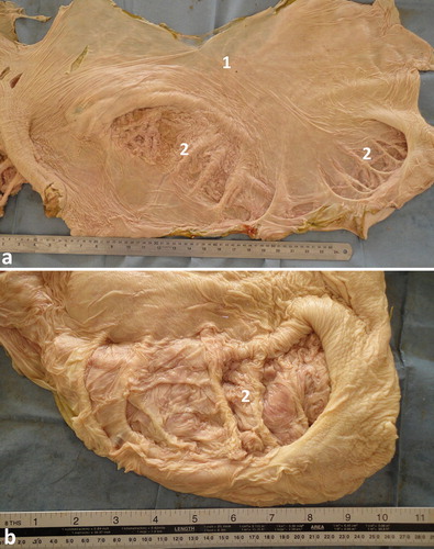
Figure 3. Anatomy of the interior of the reticulum, omasum and abomasum (a). Image (b) shows a close-up view for the rumen, reticulum and abomasum. The interior of the reticulum is characterized by a powerful and complex system of radiating pillars which are interconnected by a lattice-work of slender pillars (1). The omasum is a long sausage-shaped organ with a mucus membrane arranged in approximately 50 longitudinal folds of up to 20 mm high (2). The mucus membrane of the fundic region of the abomasum is thrown into convoluted folds parallel to the greater curvature, while the pyloric part has a smooth surface (3).
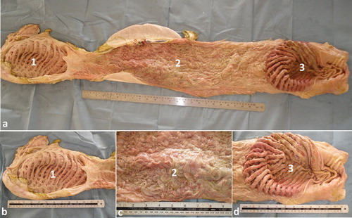
Figure 4. Anatomical position of the small and large intestine, right (a) and left (b) side view in a camel carcass preserved in 10% formalin solution. 1 = liver; 2 = right kidney; 3 = abomasum; 4 = jejunum; 5 = colon; 6 = rumen; 7 = left kidney; 8 = spleen; 9 = colon.
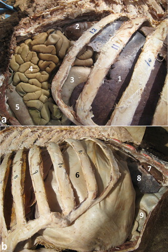
Figure 5. Ultrasonograms of the rumen and glandular sacs in a healthy camel. The rumen wall is smooth and echogenic and the contents could not be seen because of reflection of the ultrasound waves by gas contents (a). Image was taken from the upper left 10th intercostal space. Image (b) shows the glandular sacs of the cranioventral ruminal sac at the level of the 5th intercostal space on the left side. Image (c) shows the glandular sacs of the caudodorsal ruminal 10 cm cranial to the umbilicus. 1 = Ruminal wall; 2 = Rumen; D = diaphragm; S = glandular sacs; Ds = dorsal; Vt = ventral.
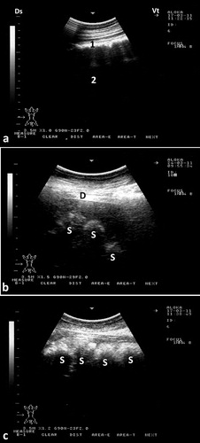
Figure 6. Ultrasonograms of the reticulum in a healthy camel. It appeared as a half-moon-shaped structure with an even contour. Images were taken at the level of the right paramedian region just behind the sternal pad. Image (a) was taken during reticular relaxation while image (b) was taken during reticular contraction. 1 = diaphragm; 2 = Reticulum; 3 = Ruminal sac; 4 = free peritoneal fluid; Ds = dorsal; Vt = ventral.
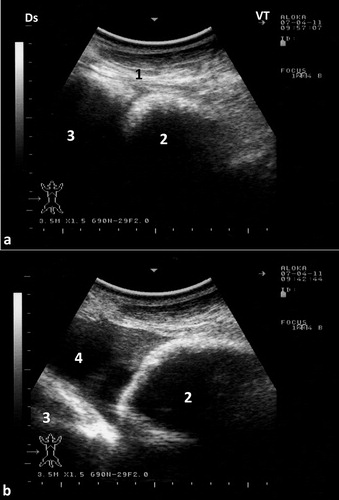
Figure 7. Ultrasonograms of the omasum and abomasum in a healthy camel. Image (a) shows the omasum and image (b) shows the abomasum at the level of the right 6th and 9th intercostal spaces, respectively. The omasal folds in image (a) appeared as fine and smooth moderately echogenic bands, while the abomasal folds in image (b) appeared as coarse and thick highly echogenic bands. 1 = liver; 2 = portal vein; 3 = omasum; 4 = abomasum. Ds = dorsal; Vt = ventral.
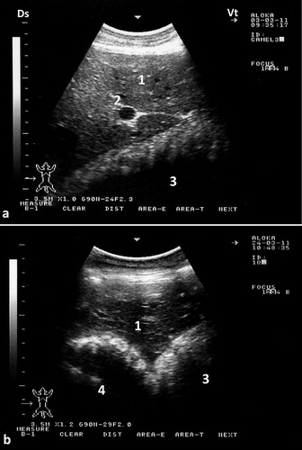
Figure 8. Ultrasonograms of the small intestine in a healthy camel. Image was taken from the ventral right paralumbar fossa. A cross sectional view was obtained in image (a) (1) while a longitudinal view was obtained in image (b) (2). Ds = dorsal; Vt = ventral.
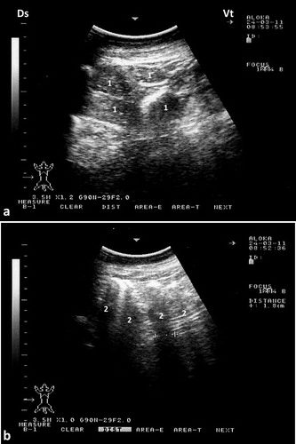
Figure 9. Ultrasonograms of the large intestine in a healthy camel. Image (a) for the cecum was taken in the caudal right flank while image (b) for the colon was taken from the anterior part of the right flank. 1 = cecum; 2 = colon; Ds = dorsal; Vt = ventral.
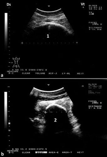
Figure 10. Aspiration of peritoneal fluid in a camel. Two sites of aspiration of peritoneal fluid were determined; anterior site is 10-cm caudal to the sternal pad (a) and posterior site is 10-cm cranial to the umbilicus (b). 1 = sternal pad; 2 = umbilicus.
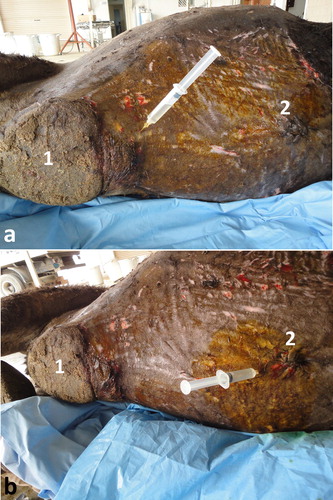
Figure 11. Ultrasound-guided abdominocentesis in a camel. Image was taken 10-cm caudal to the sternal pad. The reticulum is in a contracted position. 1: needle; 2: peritoneal fluid; 3: reticular wall. DS = dorsal; VT = ventral.
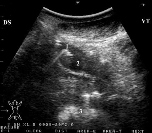
Figure 12. Ultrasonograms of intestinal obstruction in 2 camels due to foreign bodies (wool balls; FB) within the intestinal lumen (a & b). Image (c) shows a wool ball within the intestinal lumen detected at postmortem examination (white arrow). Image (d) shows multiple wool balls and other foreign bodies removed from the rumen during rumenotomy. FB = foreign body; J = jejunum.
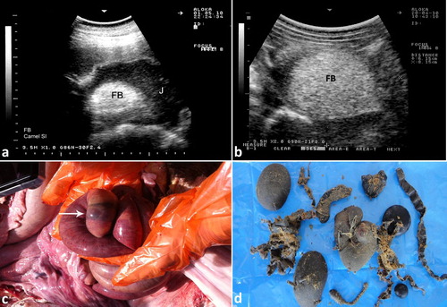
Figure 13. Intestinal volvulus in a female camel showing bilateral abdominal distension (a). Image (b) shows dilated intestinal loops with markedly reduced motility. Image (c) shows mesenteric torsion. 1 = distended intestinal loops; 2 = shows site of volvulus.
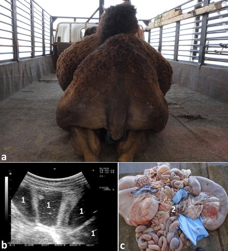
Figure 14. Ultrasonograms of intestinal intussusception in 2 camels (a & b). Image (c) shows a jejuno-jejunal intussusception in a female camel detected at postmortem examination (black arrow). Image (d) shows severe intestinal hemorrhage after jejunal separation of image (c). 1 = intussuscipiens; 2 = intussusceptum.
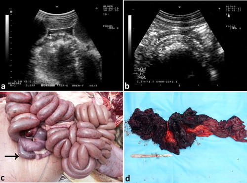
Figure 15. Ultrasonography of the rumen in a female camel admitted with colic. The ruminal contents appeared moderately echogenic (a). Image (b) shows a large amount of sand in the rumen at postmortem examination. 1 = rumen; 2 = sand; 3 = ruminal contents (barely).
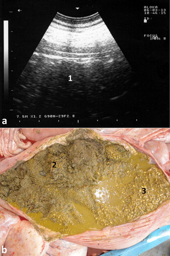
Figure 16. Severe abdominal distension in a camel with trypanosomiasis (a). Images b & c show ultrasonograms with anechoic abdominal effusions where the liver and intestines are floating. Ds = dorsal; Vt = ventral.
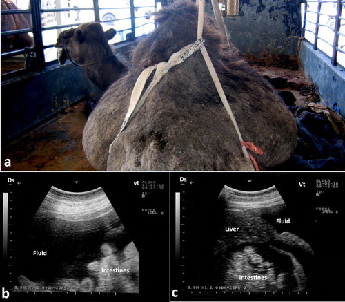
Figure 17. Chronic peritonitis in a female camel. Fibrin and hyperechoic fluid are imaged within the peritoneum exudation (a). Image (b) shows necropsy findings where massive fibrin network and adhesion of the viscera together and to the abdominal wall together with serosanguineous abdominal exudate were detected.
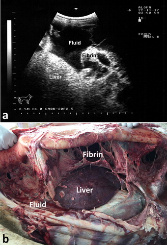
Figure 18. Pelvic abscessation in a male camel. Image with hyperechoic pattern was taken transabdominal (a). Two litres of pus was aspirated under ultrasound-guidance. Image (b) shows distended urinary bladder (UB) with hyperechogenic sedimentation.
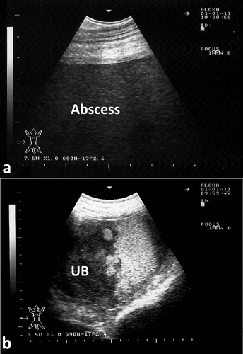
Figure 19. Necropsy findings in a female camel with difficult defecation and straining. Transrectal ultrasonography showed a pelvic mass. Necropsy was performed where abscess was detected near the uterus (a), compressing the left ureter (black arrow; b) and leading to hydronephrosis of the left kidney. Other lesions were fibrinous peritonitis and pelvic adhesions. 1 = left ovary; 2 = right ovary; 3 = uterine body; 4 = abscess.
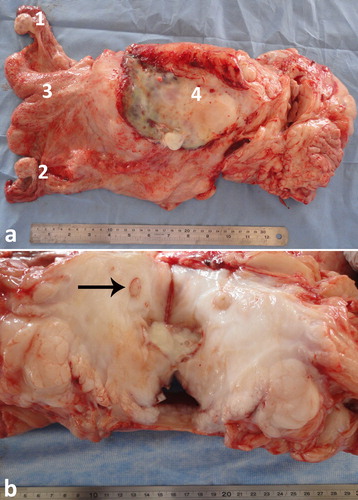
Figure 20. Paratuberculosis in a female camel with progressive weight loss (a). Image (b) shows corrugation of the intestinal mucosa (white arrows).
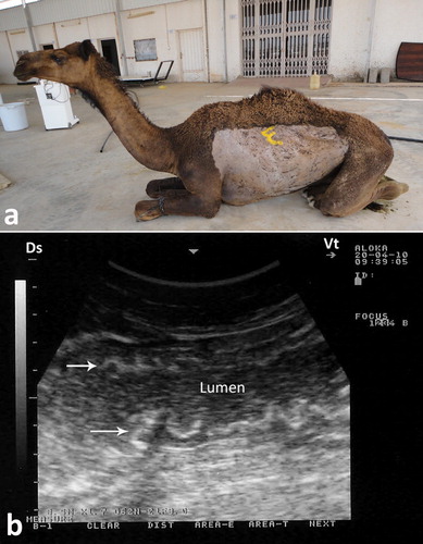
Figure 21. Paratuberculosis in a female camel with progressive weight loss (a). Image (b) shows enlargement of the mesenteric lymph nodes (1).
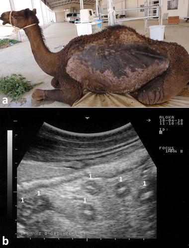
Figure 22. Necropsy finding in a female camel with paratuberculosis. Image (a) shows enlargement of the mesenteric lymph nodes (white arrows). Image (b) shows corrugation of the intestinal mucosa.
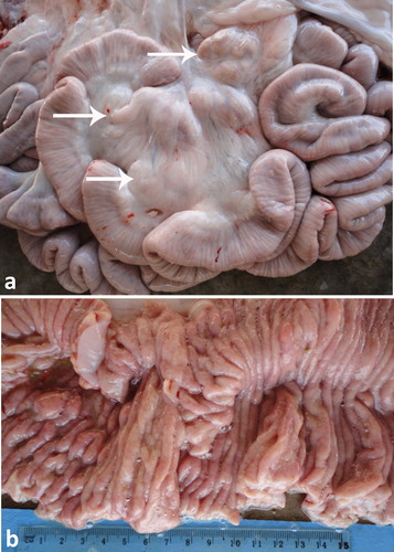
Figure 23. Intraoperative ultrasonographic view of the gastric mass showing the abomasal wall appeared thickened, vascularized, hyperechoic (white arrow, a) and the contents appeared heterogeneous (black arrow, b).
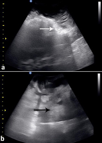
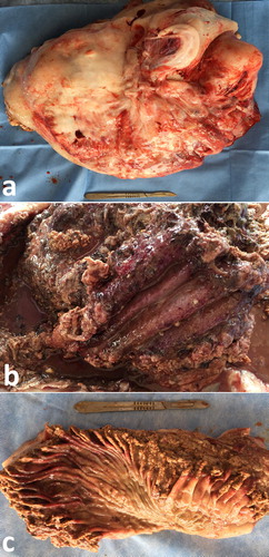
Figure 24. Abdominal enlarged hard mass weighting 10.5 kg (a). The cut section of the mass showing the abomasal (b) and omasal (c) folds were thickened, congested and ulcerated.