
Figures & data
Figure 1. Representative images of porcine blastocysts following injection of mouse induced pluripotent stem (iPS) cells: (A) blastocyst before cell injection; (B) shrunk blastocyst immediately after cell injection; (C) re-expanded blastocyst 24 h after culture; (D) hatched blastocyst 24 h after culture. Green colour represents injected iPS cells. Scale bar indicates 50 μm.
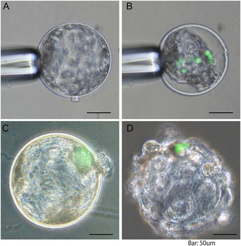
Figure 2. Effects of chlorogenic acid (CGA) and ITS supplementation during in vitro maturation on the proportions of oocytes reaching metaphase II (A) and oocytes with DNA fragmented nuclei (B), total fertilization (C), monospermic fertilization (D), blastocyst formation (E) and the total cell number of blastocysts (F). Oocytes matured in the maturation medium containing β-mercaptoethanol but not CGA and ITS served as the control group. All oocytes matured with or without CGA and ITS were used to estimate the proportions of oocytes reaching metaphase II (MII) and having nuclei with fragmented DNA (146–152 oocytes per treatment), and of fertilization (139–152 oocytes per treatment) and embryo development (199–230 oocytes per treatment). All the experiments were repeated four to five times. Each bar represents the mean value ± SEM. Bars with different letters differ significantly (P < 0.05).
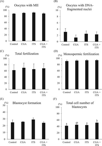
Table 1. Effects of CGA and ITS supplementation during in vitro maturation on the development of embryos following injection of mouse iPS cells into blastocysts*.