Figures & data
Figure 1. Pictures of the stages of the operation procedure.
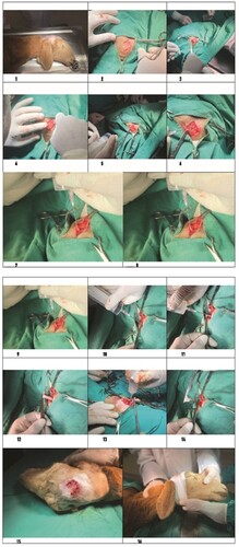
Figure 2. a: Calvarium, b: Duramater, c: Coenurus Cerabralis cyst membrane, d: Coenurus cerabralis cyst fluid, e: Brain, f: Injector, g: Extracted cyst fluid, h: Hemostatic clamp used to fix the protruding cyst part, i: stretching movements for the release of the cyst, j: the cyst is completely protruding, k: the sutured closure of the opened skin flap, l: the bandage for protection, →: the direction of movement of the cyst fluid
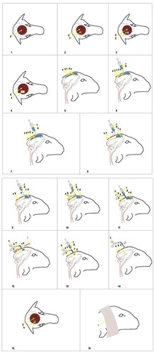
Table 1. Biochemical parameters of C. cerebralis infected sheep before and 1 month after the operation (mean ± SE).
Table 2. Hematological parameters of C. cerebralis infected sheep before and 1 month after the operation (mean ± SE).
Figure 3. Fluctuant Coenurus cerebralis cyst (arrow) filled with transparent fluid in the frontal region of the brain.

Figure 4. The cyst (arrow) in the frontal region of the brain with evacuated fluid and white scolexes.
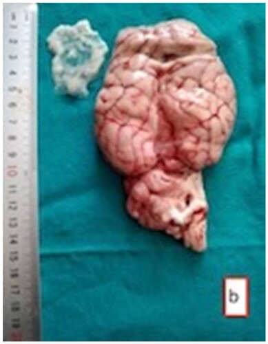
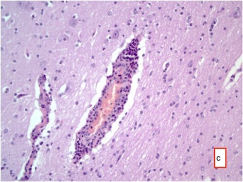
Figure 5. Cyst membrane (arrow) and mononuclear cell infiltrate in these areas with occasional eosinophil granulocytes, and perivascular mononuclear cell infiltrates HE ×200.
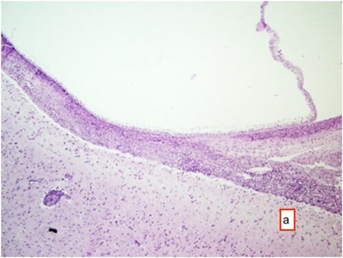
Figure 6. Mononuclear cell infiltrations in and around necrotic areas, HE ×25.
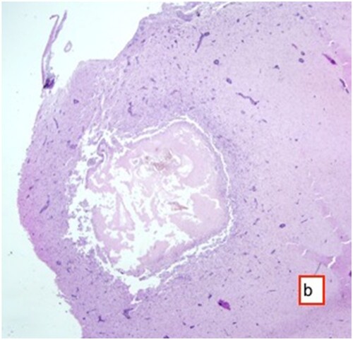
Figure 7. Hyperemia in vessels, perivascular mononuclear cell infiltrates, necrosis in some neurons and gliosis with neuronophagia, HE ×400.