
Figures & data
Table 1. Clinical score.
Figure 1. Determination of height and width of palpebral fissure, angle of the mouth, and frontal and in profile angles of the nose. A, height of palpebral fissure, straight line from the centre of the lower eyelid to the centre of the upper eyelid, intersecting the midpoint of the pupil. Width of palpebral fissure, straight line from the lateral to the medial corner of the right eye, crossing the middle of the pupil. Profile nose angle, one axis was placed over the right nostril, and the other axis was aligned to touch the most rostral point of the nose. The connecting vertex was then situated at the most ventral point of the nose. Mouth angle, one axis parallel to the oral commissure, while the other axis was drawn sagittally from the frontal bone at the level of the supraorbital foramina, tangentially touching the tip of the nose. The connecting vertex was positioned in front of the upper lip. B, frontal angle of the nose, two axes were strategically positioned over each nostril, with the connecting vertex situated at the convergence point of the nostrils.
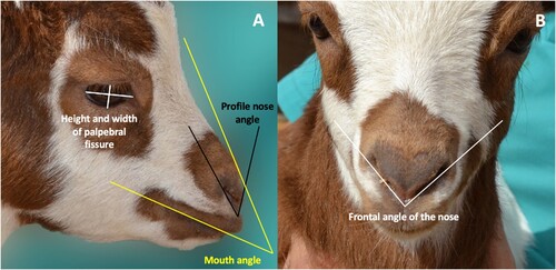
Table 2. Descriptive statistics of frontal angle of the nose in degrees.
Table 3. Descriptive statistics for height and width of palpebral fissure, angle of the mouth and in profile angle of the nose.
Table 4. Least square means of mouth angle and side nose angle according to pain classification.
Table 5. Kruskal–Wallis analysis and post hoc Wilcoxon rank for Frontal angle of the nose.