Figures & data
Figure 1 Flow diagram of selection process in the meta-analysis.
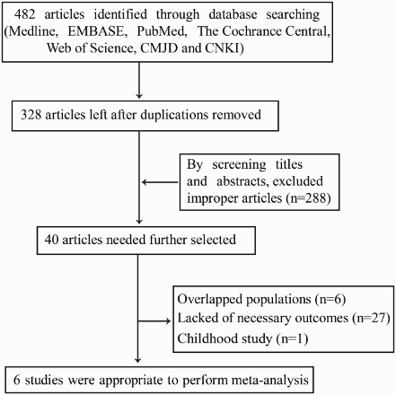
Table 1 Summary of the data extracted from the six studies included
Table 2 Determination of IPSS risk groups
Table 3 Combined mutations of ASXL1 with IDH1/2, NPM1, RUNX1, and TET2 in MDSs
Table 4 Quality assessment of individual study
Figure 2 Forest plots of the HRs and 95% CIs for OS and LFS in FAB-defined MDS patients. The size of the blocks or diamonds represents the weight and the length of the straight line represents the width of 95% CI.
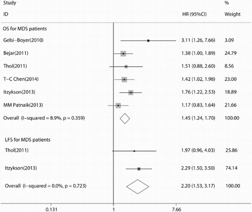
Figure 3 Begg's funnel plot with pseudo 95% confidence limits.
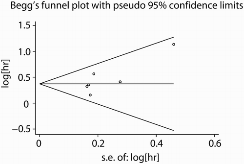
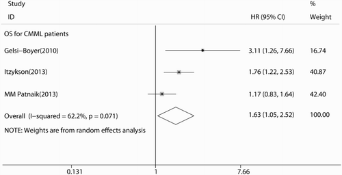
Figure 4 Forest plots of the HRs and 95% CIs for: OS in CMML patients. The size of the blocks or diamonds represents the weight and the length of the straight line represents the width of 95% CI.