
Figures & data
Figure 1 (A) Dysplastic BM erythropoiesis; intranuclear inclusion giant pronormoblasts (Giemsa). (B) Immunhistochemical contrasting of erythropoiesis: abnormal BM dysplasia, no aplasia. (C) Immunohistochemical detection of PVB19-positive cells in BM (antiPVB19 Ab)
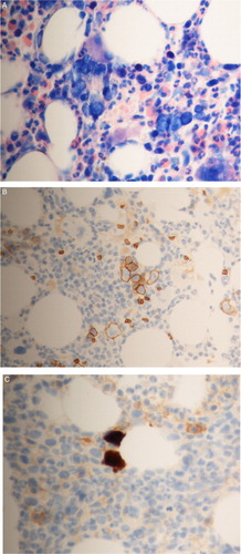
Figure 2 Correlation of PVB19 diagnostic parameters (PVB19 viral load, lgG/lgM immunoblot) with anemia laboratory values (reticulocytes, hemoglobin) over time during treatment (prednisolone, MMF, IVIG)
Figure 3 PVB19 immunoblot (recomline®): line 1: PVB19-IgG-immunoblot (positive): Vp-2p +++, VP-1S +, VP-2r [+]; line 2: PVB19-IgG avidity: (VP-1S); line 3: PVB19 IgM-immunoblot (positive): Vp-2p +++, VP-2r +, Vp-C [+]. Abbreviations: Vp-2p (main capsid antigen- conformation epitope), VP-1S (VP1-unique region), VP-2r (main capsid antigen- linear epitope), Vp-C (C – terminal conjoint half of structure proteins VP1 and VP2).
![Figure 3 PVB19 immunoblot (recomline®): line 1: PVB19-IgG-immunoblot (positive): Vp-2p +++, VP-1S +, VP-2r [+]; line 2: PVB19-IgG avidity: (VP-1S); line 3: PVB19 IgM-immunoblot (positive): Vp-2p +++, VP-2r +, Vp-C [+]. Abbreviations: Vp-2p (main capsid antigen- conformation epitope), VP-1S (VP1-unique region), VP-2r (main capsid antigen- linear epitope), Vp-C (C – terminal conjoint half of structure proteins VP1 and VP2).](/cms/asset/dda858b3-1eaf-4614-bf98-7a40e8da13cc/yhem_a_1183288_f0003_c.jpg)