Figures & data
Figure 1 Mechanisms of cellular and humoral immune response: the role of T and B lymphocytes.
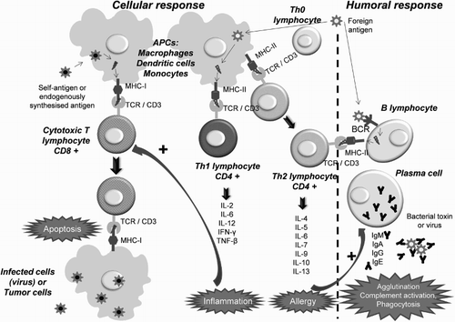
Figure 2 Immunological alterations in ITP: (A) the presence of an inflammatory stimulus or a stressful situation (associated with an infection, drugs, etc.) transforms certain platelet GPs into autoantigens that trigger an immune response; (B) antigen-presenting cells (activated macrophages) process the platelet autoantigens and present them on MHC class I and II molecules; (C) circulating Th cells with TCR specific for platelet antigens activate and become autoreactive, secreting cytokines which act as co-stimulatory signals of B lymphocytes; (D) activated autoreactive B cells recognize platelet GPs and initiate mass production of autoantibodies (IgG) that destroy platelets and megakaryocytes through the humoral response; (E) in parallel, activated macrophages present platelet autoantigens to cytotoxic T cells and activate them, inducing platelet destruction mediated by the cellular response. Adapted from John W. Semple's Laboratory Homepage, by M. Perera. Retrieved from http://www.angelfire.com/ut/johnsnotes/index.html. Copyright 2009 by John W. Semple. Adapted with permission.
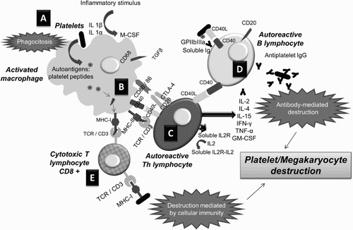
Table 1 Main alterations of the immune system described in patients with ITP and the possible effects achievable using different available therapies
Figure 3 Possible role of platelet TLRs in the pathophysiology of ITP: platelets act as circulating sentinels and, in the event of detecting pathogens or molecules derived from injured tissues, the TLRs that recognize them send ‘danger signals’ to the immune system by stimulating the effector cells and the APCs of the RES; if anti-platelet antibodies are also present, the TLRs stimulate R-Fc-mediated phagocytosis. Adapted from John W. Semple's Laboratory Homepage, by M. Perera. Retrieved from http://www.angelfire.com/ut/johnsnotes/index.html. Copyright 2009 by John W. Semple. Adapted with permission.
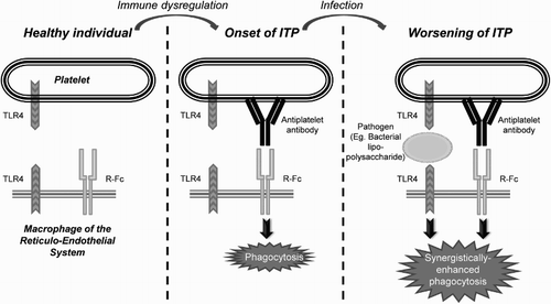
Figure 4 Alterations in T lymphocytes in active ITP (pro-inflammatory state): there is an increase in Th17 cells and a decrease in Treg levels, which release less amount of the anti-inflammatory cytokine TGF-β to the blood. These changes are reversed after the response to several treatments, such as A-TPOs. Adapted by M. Perera with permission from John W. Semple. Copyright 2009 by John W. Semple.
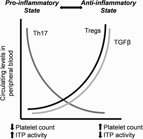
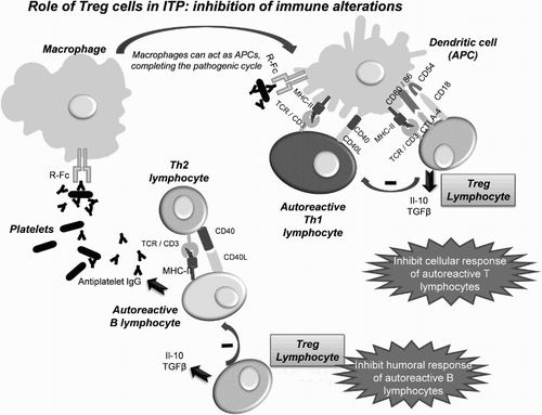
Figure 5 Effects of recovery in the level of Treg cells in ITP patients: the Tregs inhibit the activity of autoreactive effector T cells (CTL and Th) in the presence of APCs with autoantigens by releasing soluble IL-10 and TGF-β; they also inhibit, by direct contact, autoreactive B lymphocytes, which fail to produce antibodies against platelets.