Figures & data
Table 1. Patient characteristics.
Figure 1. Kinetics of T-cell subsets and sjTREC after AHSCT. In each panel, results are expressed as absolute number of cells per μl periphery blood for T-cell subsets, copies per 106 PBMCs and copies per ml periphery blood for sjTREC. Results of healthy donors are also included in each panel. Different panels show the distribution of a distinct cell subset at different time points: before AHSCT, and at 1 month, 2 months, 3 months. (A) CD3+T cells. (B) CD3+CD8+T cells. (C) CD3+CD4+T cells. (D) CD4+CD45RA+CD31+T cells. (E) sjTREC per ml blood. (F) sjTREC per 106 PBMCs. (G) CD4+CD45RA+CD62L+T cells. Each box shows the median, quartiles, and extreme values.
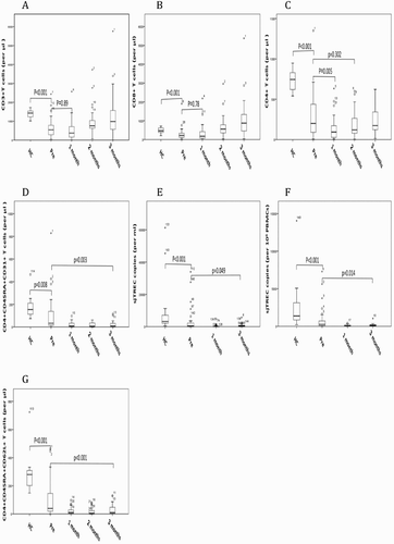
Figure 2. sjTREC level was of predictive value for aGVHD in patients after AHSCT. Cumulative incidence of aGVHD in patients who underwent AHSCT (n = 40). Gray line indicates high sjTREC level, black dashed line indicates low sjTREC level. sjTREC levels are expressed in sjTREC per 106 PBMCs (A) and sjTREC per ml blood (B).
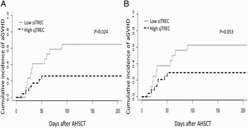
Table 2. Cox multivariate analysis.
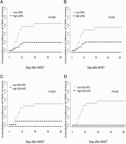
Figure 3. sjTREC level and CD31+RTE were of predictive value for CMV viremia in patients after AHSCT. Cumulative incidence of CMV viremia in patients who underwent AHSCT. Gray line indicates high sjTREC level, black dashed line indicates low sjTREC level. sjTREC are expressed in sjTREC copies per 106 PBMCs (n = 40) (A) and sjTREC copies per ml blood (n = 40) (B) CD31+RTE are expressed in percentage in CD4+T cells (n = 32) (C) and μl blood (n = 31) (D).