Figures & data
Figure 1. Hemoglobin analysis of the two patients (P1 and P2) who were respectively compound heterozygotes for Hb G-Georgia/α0-thalassemia and Hb Nakhon Ratchasima/α0-thalassemia using automated HPLC and CE systems. The separated profiles of Hb G-Georgia, Hb Nakhon Ratchasima (NR), Hb A, Hb A2 and Hb A2NR are depicted.
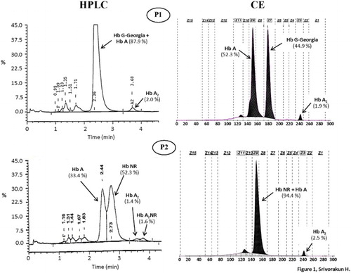
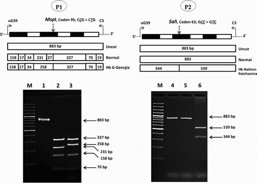
Figure 2. Identification of the Hb G-Georgia and Hb Nakhon Ratchasima mutations by PCR-RFLP assays using MspI and SalI digestion, respectively. The locations and orientations of primers αG39 and C3 and the size of amplified α2-globin gene fragment (883 bp) are illustrated. The sizes of MspI and SalI-digested fragments specific for αG-Georgia (258 bp) and αNakhon Ratchasima (539 bp and 344 bp) and their normal counterparts are indicated. In agarose gel electrophoresis, M represents the GeneRuler 50 bp DNA ladders. 1 and 4: undigested amplified DNA, 2: MspI-digested amplified DNA of normal subject, 3: MspI-digested amplified DNA of the P1 with Hb G-Georgia/α0-thalassemia, 5: SalI-digested amplified DNA of normal subject, 6: SalI-digested amplified DNA of the P2 with Hb Nakhon Ratchasima/α0-thalassemia.