Figures & data
Table 1. Clinical characteristics of MM patients and normal donors.
Figure 1. The proliferation and apoptosis of RPMI-8226 cells transfected with shRNA-REGγ-1 and shRNA-REGγ-2 (n = 3). (A) The proliferation rates of RPMI-8226 cells transfected with shRNA-REGγ-1 and shRNA-REGγ-2 by CCK8. (B) The apoptosis of RPMI-8226 cells transfected with shRNA-REGγ-1 and shRNA-REGγ-2 by flow cytometry. (C) The apoptosis rates of RPMI-8226 cells transfected with shRNA-REGγ-1 and shRNA-REGγ-2. *Represent significantly different at p < 0.05 when compared with the control (RPMI-8226 cells transfected with shN).
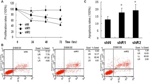
Figure 2. The migration of RPMI-8226 cells transfected with shRNA-REGγ-1 and shRNA-REGγ-2 by transwell assay (n = 3). (A) Microscopic observation of migrated RPMI-8226 cells. (B) The number of migrated cells. **Represent significantly different at p < 0.01 when compared with the control (RPMI-8226 cells transfected with shN).
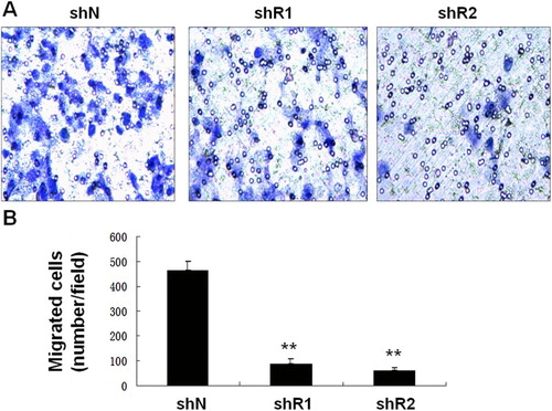
Figure 3. The potential regulatory mechanisms of REGγ on RPMI-8226 cells. (A) The expression of p-p65, p65, IkBε and αREGγ in RPMI-8226 cells transfected with shRNA-REGγ-1 and shRNA-REGγ-2 by Western blot (n = 3). (B) The expression of IkBε in RPMI-8226 cells transfected with shRNA-REGγ-1 and shRNA-REGγ-2 by RT-PCR. (C) The degradation of IkBε (half-life period) in RPMI-8226 cells transfected with shRNA-REGγ-1. (D) The expression of REGγ, MMP2, Bcl-xL and XIAP in RPMI-8226 cells transfected with shRNA-REGγ-1 and shRNA-REGγ-2 by Western blot.