Figures & data
Figure 1. The effect of TFP on ALK+ ALCL cell viability. (A) Cell viability of DEL cells with or without TFP treatment at 24 and 48 hours. (B) Cell viability of SUP-M2 cells with or without TFP treatment at 24 and 48 hours. (C) Cell viability of human normal lymphocyte cells with TFP or without TFP treatment at 24 and 48 hours. Cell survival was determined by TB assay. The experiments were repeated three times with consistent results. *p < 0.05. **p < 0.01.
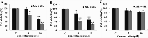
Figure 2. Cell cycle arrest and apoptosis induced by TFP. (A, B) Cell cycle distribution of DEL and SUP-M2 cells with or without TFP treatment at 24 and 48 hours. The two cell lines after treated with TFP exhibited cell proportions in the G0/G1 phase increased significantly at 48 hours while no change at 24 hours. Representative results of three separate experiments are shown. (C, D) Cell apoptosis was assessed by Annexin V-FITC/PI Apoptosis Detection Kit. The average percent of AnnexinV-FTIC positive cells is shown to increase gradually in time- and concentration-dependent manners. *Significant difference at p < 0.05. **Extremely significant difference at p < 0.01. The experiments were repeated three times.
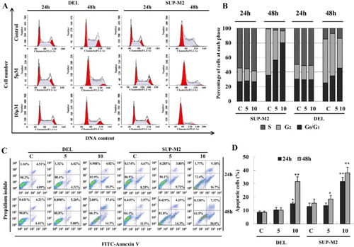
Figure 3. Effects of TFP on cell cycle and apoptosis regulatory molecules. (A, B) Cells were harvested at indicated time points for RT-PCR and Q-PCR reactions. Both p21 and p27 mRNA levels were increased significantly while CDK2, CDK4 and Cyclin E1 were decreased at 24 hours after treatment with TFP. The data represent results from one of three independent experiments. (C) Western Blot of cell cycle checkpoint proteins after treatment with TFP. P21 and p27 protein levels were increased at both 24 and 48 hours, whereas CDK2 was decreased remarkably at 48 hours. (D) Western blot of apoptosis-related proteins. Bax demonstrated a relatively stable expression while Bcl-XL was decreased continually at both 24 and 48 hours, and Bcl-2 was reduced to a lower level at 24 hours but was increased significantly at 48 hours. (E) Western blot of NPM-ALK and related downstream survival proteins after treatment with TFP. At 24 and 48 hours, NPM-ALK was degraded while phosphorylated NPM-ALK showed an elevation. Phospho-AKT had no change with phospho-ERK was increased and phospho-STAT3 was decreased at 24 hours. At 48 hours, phospho-ERK, AKT and STAT3 were increased significantly.
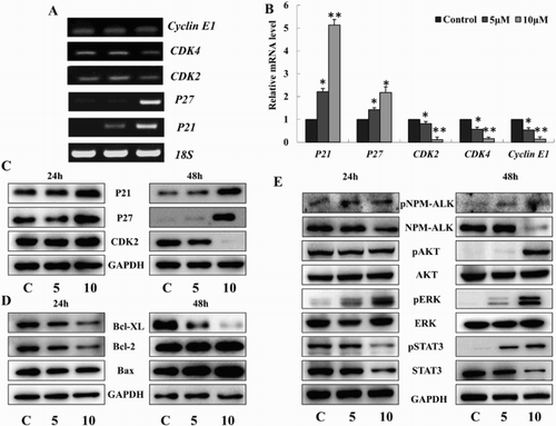
Figure 4. Decreased tyrosine kinase activity of NPM-ALK induced by TFP. Tyrosine kinase activity of NPM-ALK shows a gradual decrease after treatment of TFP for 48 hours. *Significant difference at p < 0.05. The experiments were repeated three times.
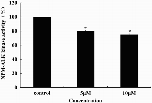
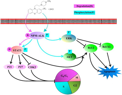
Figure 5. The mode of action of TFP in ALK+ ALCL cells. TFP inhibited the cell viability via induction of the degradation and decrease of tyrosine kinase activity of NPM-ALK. The cell apoptosis occurs through the activation of ERK pathway while G0/G1 cell cycle was arrested through the degradation of STAT3.