Figures & data
Table 1. Maternal and fetal blood gases before brain collection.
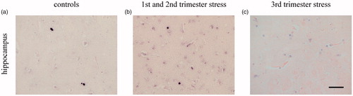
Figure 1. Effects of chronic maternal stress on neuronal network formation in fetal sheep brain at 0.87 gestation. Silver staining of the cerebral cortex (cor), superficial white matter (sfwm), subcortical white matter (swm), deep white matter (dwm) and the CA3 region of the hippocampus (hc). *p<.05, compared to controls; $$p<.01 compared to the next more superficial brain region; §§p<.01 compared to the hippocampus. Controls: n = 8, 1st/2nd trimester stress: n = 10, 3rd trimester stress: n = 10.
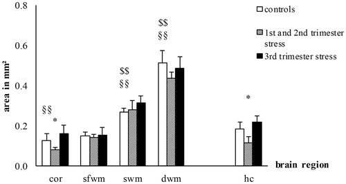
Figure 2. Effects of chronic maternal stress on neuronal network formation in frontal cortex and hippocampus of the fetal sheep brain at 0.87 gestation. Representative photomicrographs of silver staining of the cerebral cortex (a–c) and the CA3 region of the hippocampus (d–f). Stress during the first and second trimester but not stress during the third trimester reduced the area of silver staining. Scale bar 50 µm.
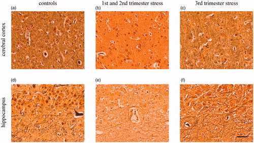
Table 2. Chronic effects of maternal psychosocial stress on structural brain development at 0.87 gestation.
Figure 3. Effects of chronic maternal stress on synaptic density in fetal sheep brain at 0.87 gestation. Anti-synaptophysin-immunohistochemistry in the cerebral cortex and the CA3 region of the hippocampus. *p<.05, **p<.01 compared to controls; §§p<.01 compared to the hippocampus. Controls: n = 8, 1st/2nd trimester stress: n = 10, 3rd trimester stress: n = 10.
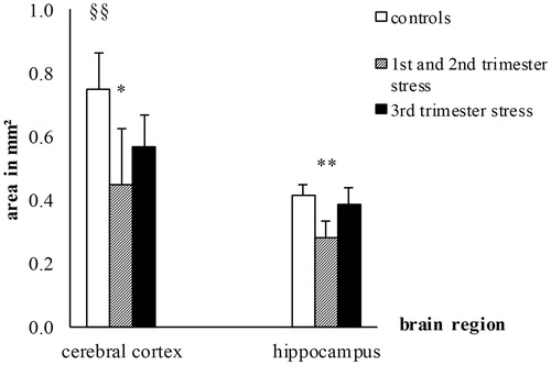
Figure 4. Effects of chronic maternal stress on synaptic density in frontal cortex of the fetal sheep brain at 0.87 gestation. Representative photomicrographs of synaptophysin immunohistochemistry (brown precipitation) of the CA3 region of the hippocampus (a–c). Stress during the first and second trimester but not stress during the third trimester reduced synaptophysin IR. Scale bar 100 µm.
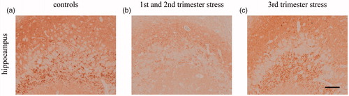
Figure 5. Effects of chronic maternal stress on myelination in fetal sheep brain at 0.87 gestation. Anti-MBP-immunohistochemistry of the cerebral cortex (cor), superficial white matter (sfwm), subcortical white matter (swm), deep white matter (dwm), and the CA3 region of the hippocampus (hc). *p<.05, **p<.01 compared to controls; $$p<.01 compared to the next more superficial brain region; §§p<.01 compared to the hippocampus. Controls: n = 8, 1st/2nd trimester stress: n = 10, 3rd trimester stress: n = 10.

Figure 6. Effects of chronic maternal stress on myelination in white matter of fetal sheep brain at 0.87 gestation. Representative photomicrographs of MBP immunohistochemistry (brown precipitation) of the superficial white matter (a–c) and the subcortical white matter (d–f). Stress during the first and second trimester but not stress during the third trimester reduced MBP IR. Scale bar 200 µm.
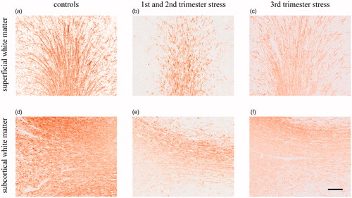
Figure 7. Effects of chronic maternal stress on cell proliferation in fetal sheep brain at 0.87 gestation. Anti-ki-67-immunohistochemistry of the cerebral cortex (cor), superficial white matter (sfwm), subcortical white matter (swm), deep white matter (dwm) and the CA3 region of the hippocampus (hc). *p<.05 compared to controls; $p<.05 compared to the next more superficial brain region. Controls: n = 8, 1st/2nd trimester stress: n = 10, 3rd trimester stress: n = 10.
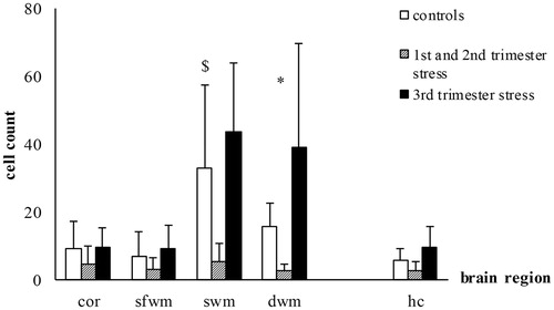
Figure 8. Effects of chronic maternal stress on cell proliferation in white matter of fetal sheep brain at 0.87 gestation. Representative photomicrographs of cell proliferation (ki-67 immunohistochemistry, brown precipitation) of the deep white matter (a–c). Stress during the first and second trimester but not stress during the third trimester decreased cell proliferation seen by the lower number of ki-67 marked cells. Scale bar 50 µm.
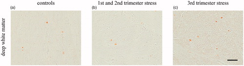
Figure 9. Effects of chronic maternal stress on developmental cell death in fetal sheep brain at 0.87 gestation. TUNEL-method of the cerebral cortex (cor), superficial white matter (sfwm), subcortical white matter (swm), deep white matter (dwm) and the CA3 region of the hippocampus (hc). Controls: n = 8, 1st/2nd trimester stress: n = 10, 3rd trimester stress: n = 10.
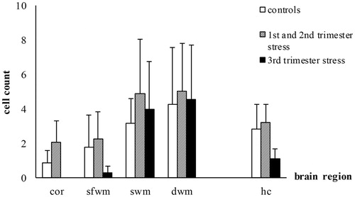
Figure 10. Effects of chronic maternal stress on developmental cell death in white matter of fetal sheep brain at 0.87 gestation. Representative photomicrographs of developmental cell death (TUNEL, black color, counterstaining with Nuclear Fast Red) of the deep white matter (a–c). Partly due to the low number of TUNEL marked cells and the high variance there were no effects on developmental cell death. Scale bar 50 µm.