Figures & data
Figure 1. ELS reduces the number of β-tanycytes in the hypothalamus of adult male mice. The ELS paradigm consisting of limited nesting/bedding-material results in significant reductions in body weight (A) and body weight gain between P2 and P9 (B). In the hypothalamus, tanycytes were co-labeled with GFP and SOX2 (C). Nuclear DNA was labeled with DAPI. α-tanycytes and β-tanycytes were quantified in the hypothalamic areas outlined dashed lines. Adult mice exposed to ELS between P2 and P9 showed a significant decrease in the number of β-tanycytes (E), while no differences in the numbers of α-tanycytes were observed (D). BW: body weight; *p < 0.05.
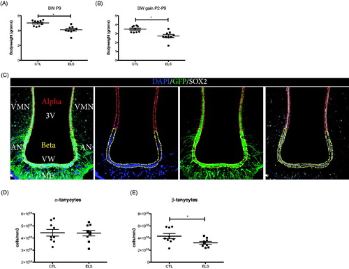
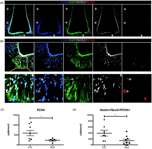
Figure 2. ELS reduces cell proliferation in the hypothalamus of adult male mice. Proliferating (PCNA+) cells, were found in parenchymal areas surrounding the 3rd ventricle but not on the ventricular wall of the hypothalamus in Nestin-GFP mice (A). Representative image from a male control mouse. PCNA + cells were found in the parenchyma (B) and there, total numbers of proliferating PCNA + cells and putative proliferating NSCs positive for Nestin-GFP, SOX2 and PCNA in the ME were quantified. ELS resulted in a significant reduction of the total numbers of PCNA + cells (C) and specifically in a significant reduction of PCNA+/SOX2+/Nestin + proliferative cells (D) in the ME. *p < 0.05.