Figures & data
Figure 1. CUMS rat model was successfully established. Tail suspension (A), Sucrose preference (B), and Forced swimming (C) in rats with or without exposure to CUMS. The sugar and water preference rate of rats in the stress group decreased significantly (p < 0.01), and the immobility time in the tail suspension experiment as well as the forced swimming experiment increased significantly. The concentration of ACTH (D,F) and CORT (E,G) in the serum of CUMS model rats increased significantly before and after the orthodontic application *p < 0.05, **p < 0.01, ***p < 0.001, compared with the control group.

Figure 2. Orthodontic tooth movement can alter the bone metabolism balance changes. (A) The tooth movement distance of rats in the stress group was markedly increased. The concentration of CTX (C) in the serum was increased but the concentration of PINP (B) was decreased. These results suggest that the osteoclast activity of rats in the stress group increased, but the osteogenic activity decreased. The balance of bone metabolism was disrupted. *p < 0.05, **p < 0.01, ***p < 0.001, compared with the control group.
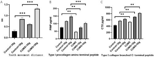
Figure 3. HE staining showed that the alveolar bone fibers in the tooth movement area in the CUMS group were sparse and osteoclast lacuna was substantially increased.
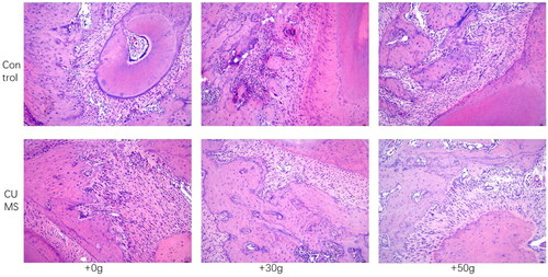
Figure 4. TRAP staining revealed increased staining of osteoclasts in the alveolar bone in the tooth movement area in the CUMS group.
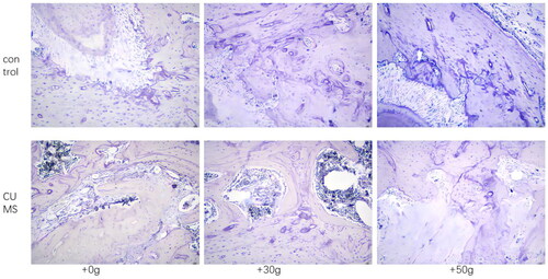
Figure 5. Before exposure (A) and after exposure (B) to orthodontic stress, the PTH content in the CUMS group was found to be significantly increased. *p < 0.05, **p < 0.01, ***p < 0.001, compared with the control group.
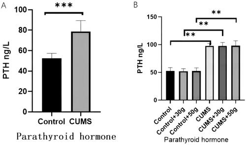
Figure 6. The AC content in the jaw bone tissue of the CUMS group did not change significantly, but the PLC content was significantly increased.
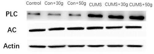
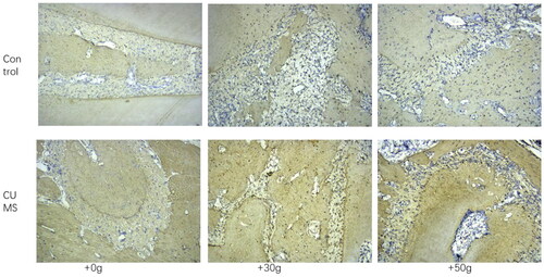
Figure 7. The expression of NF-ĸB in the jaw bone of orthodontic tooth movement area was substntially increased in CUMS group.