Figures & data
Table 1. The ligands of mineralocorticoid receptor.
Figure 1. Schematic representation of intracellular genomic and non-genomic signaling pathways following MR ligand binding. Rapid MR signaling via non-genomic pathways activates downstream receptor kinases and second messenger systems, ultimately leading to alterations in neuronal activity. Conversely, for the genomic pathway, ligand-activated MR dissociates from its chaperone protein complex, translocates to the nucleus and regulates gene transcription of HRE-containing target cells. In 11ß-HSD2 containing cells the main natural ligand of MR is aldosterone, as the high-affinity ligand cortisol (or corticosterone) will be converted. In cells not containing 11ß-HSD2, as for example many neurons, the main ligand for MR is cortisol or corticosterone. MR: mineralocorticoid receptor; HRE: hormone response element; RAS: renin-angiotensin system; ERK: extracellular signal-regulated kinase; PI3K: Phosphatidylinositide 3-kinases; Akt: protein kinase B; PKC: protein kinases C; PKD: protein kinases D. (Illustration is created with Biorender.com).
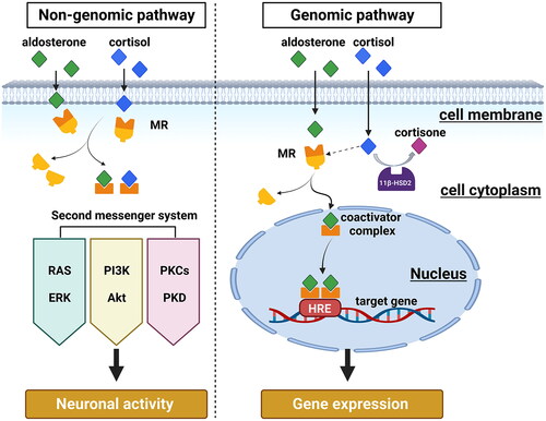
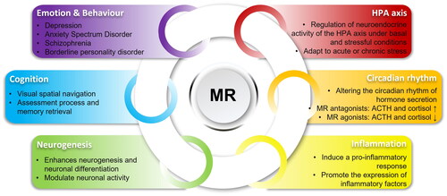
Figure 2. Overview of MR functions in the brain. In the brain, MR regulates the HPA axis activity and circadian rhythm, modulates neurogenesis, participates in neuroinflammation and affects mood, behavior, and cognition.